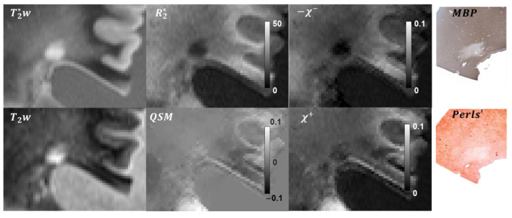
Figure 2.
Example of the -based separation of magnetic sources in a chronic silent lesion. The lesion appears to be weakly paramagnetic in the susceptibility map, with the Perls’ and MBP staining suggesting almost complete loss of myelin and partial loss of iron within the lesion ROI. These findings were similarly reflected in the estimated and maps.

