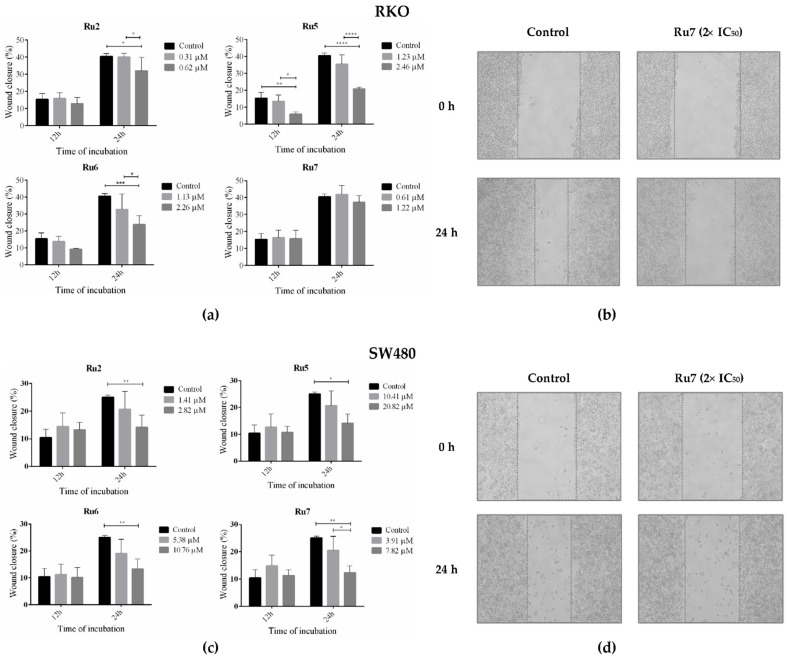
Figure 9.
Quantitative analyses of wound closure (%) the RKO (a) and SW480 (c) cell lines incubated for 12 and 24 h with IC50 and 2× IC50 with Ru2, 5–7. Wound closure was calculated from the area in time 0 h. Representative images (100×) of the wound-healing assay of the RKO (b) and SW480 (d) cell lines after incubation with 2× IC50 for 0 and 24 h. Data are presented as mean ± SD from at least three independent experiments. Results were statistically different from the negative control for * p < 0.05, ** p < 0.01, *** p < 0.001 and **** p < 0.0001.

