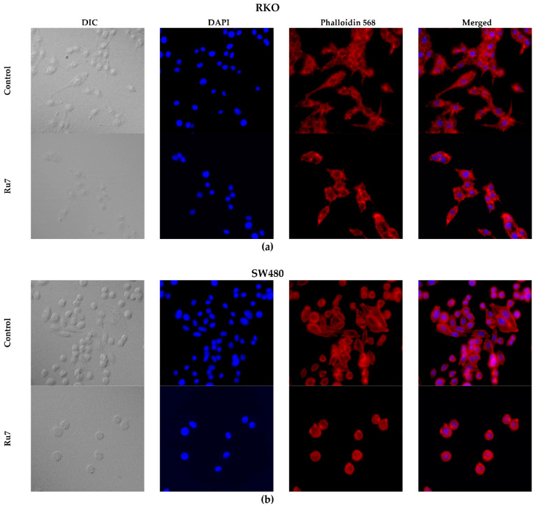
Figure 10.
Morphological changes in the cytoskeletons of the RKO (a) and SW480 (b) cells after incubation with Ru7 for 48 h. Representative images (400×) were obtained in a fluorescence microscope. Pictures of differential interference contrast (DIC), nuclei stained with DAPI (blue), F-actin-stained with Phalloidin-Alexa Fluor® 568 (red) and merged. Similar effects were found with the remaining complexes (data not shown).

