Figure 3.
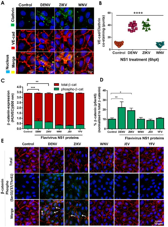
Treatment of HUVEC monolayers with DENV and ZIKV NS1 proteins but not WNV NS1 increases the colocalization of VE-cadherin with clathrin and the phosphorylation and cytosolic accumulation of β-catenin. Confluent monolayers of HUVEC were individually treated with five different NS1 proteins from DENV, ZIKV, WNV, JEV and YFV (5 μg/mL), and 6 hpt, cell monolayers were processed by (A,B) IFA for co-staining of VE-cadherin and clathrin heavy chain, (C,D) commercial ELISA for detection of total/phosphorylated β-catenin (S45), or (E) IFA for co-staining of total β-catenin (red) and phosphorylated β-catenin (S33, S37, T41) (green). Images are representative of three independent experiments. White arrows indicate VE-cadherin co-localization with clathrin heavy chain proteins (A) and the cytosolic accumulation of phosphorylated β-catenin (green) (E). Merge is indicated in yellow. IFA co-staining analyses were performed by using RBG images and ImageJ software analysis. For both assays, untreated (no NS1) cells were used as a control for normal β-catenin expression/distribution and phosphorylation in HUVEC monolayers. Images are representative of three independent experiments (area: 29.16 mm2). White arrowheads indicate colocalizing puncta of VE-cadherin and clathrin proteins. Scale bar: 10 μm. Magnification: 20×. Significant differences as follows: p < 0.05 *, p < 0.01 **, p < 0.001 ***, p < 0.0001 ****.

