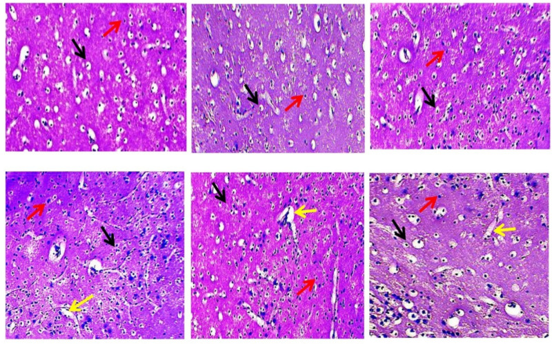
Figure 18.
The effect of 4-hydroxyisoleucine on histopathological alterations in rats following methyl mercury exposure. The slides were selected to show neuronal populations stained with hematoxylin and eosin under a fluorescence microscope. Light microphotographs of the cerebral cortex revealed the following morphological changes during MeHg exposure and 4-HI treatment: ‘Section: A’ shows the vehicle group.The black arrows indicate healthy oligodendrocyte cells, mostly found in the cerebral cortex. The red arrow indicates microglia. ‘Section: B’ shows the sham control group, with black arrows indicating healthy oligodendrocyte cells and red arrows signifying microglia. ‘Section: C’ shows the 4-HI-perse-treated group, which exhibits the typical pattern of all neuronal cells, just like the sham control group. ‘Section: D’ depicts a MeHg-treated group with oligodendrocyte degeneration and necrosis, as indicated by the black and yellow arrows, as well as an increase in microglial density (red arrow). ‘Section: E’ represents theMeHg- and 4-HI-(50 mg/kg)-treated group showing regeneration of oligodendrocytes represented by the black arrow, decreased density of microglial cells represented by the red arrow, and recovery of neuronal cells by decreasing the area of necrosis represented by the yellow arrow. ‘Section: F’ depicts the diminished area of necrosis indicated by the yellow arrow in the MeHg- and 4-HI-(100 mg/kg)-treated group, the black arrow represents the restoration of oligodendrocyte, and the red arrow represents reduced microglial density.

