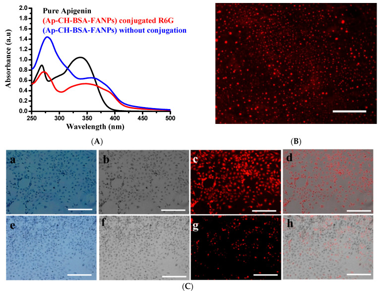
Figure 7.
UV visible of R6G conjugated to Ap-CH-BSA-NPs or Ap-CH-BSA-FANPs (A). Fluorescence image of Ap-CH-BSA-FANPs conjugated R6G (B). Qualitative cell internalization assay using R6G conjugated different nanoparticles using fluorescence microscopy (C). Fluorescence images demonstrate cellular internalization of Ap-CH-BSA-FANPs conjugated R6G in HePG-2 cells; (a) crystal violet to show cell morphology. (b) grayscale image of crystal violet. (c) TRIC channel of R6G (d) merges between grayscale image and TRIC channel by using Image j program. Fluorescence images demonstrate cellular internalization of Ap-CH-BSA-NPs conjugated R6G in HePG-2 cells (e) crystal violet to show cell morphology. (f) grayscale image of crystal violet. (g) TRIC channel of R6G (h) merges between grayscale image and TRIC channel by using Image j program.

