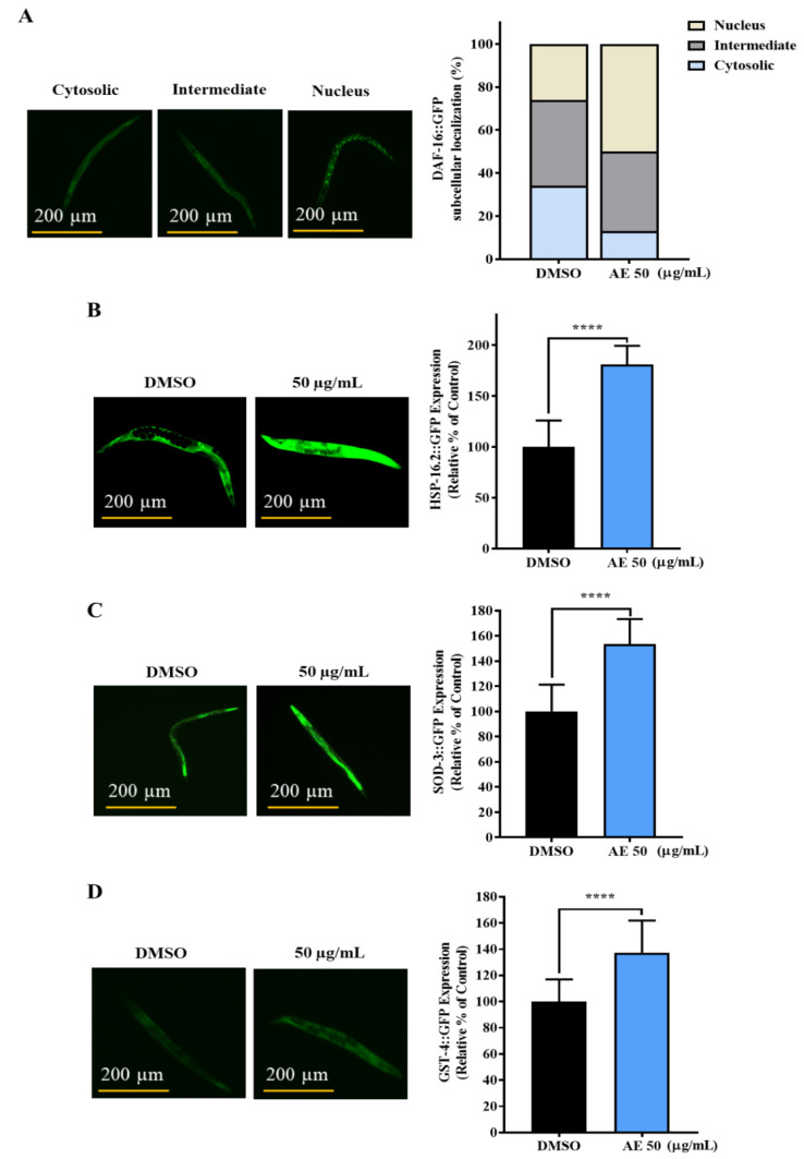
Figure 4.
Effects of AE on DAF-16 cellular translocation and the expression of HSP-16.2, SOD-3 and GST-4 in GFP-tagged worms. (A) Subcellular localization of DAF-16::GFP worms treated with or without AE (50 µg/mL) for 96 h (n = 30 worms/group). Subcellular localization was categorized into Cytosolic, Nucleus and Intermediate (between cytosolic and nucleus). (B) HSP-16.2::GFP worms treated or not treated with AE were cultured for 72 h and exposed to heat shock at 35 °C for 2 h (n = 30 worms/group). (C) SOD-3::GFP and (D) GST-4::GFP AE-treated and non-treated worms were cultured for 72 h (n = 30 worms/group). Statistical differences compared to control (DMSO) were considered significant at **** p < 0.001 by Student’s t-test. Data were represented by mean ± SD. Experiments were performed in triplicate determinations and representatives shown. Scale bar for GFP analysis = 200 µm.

