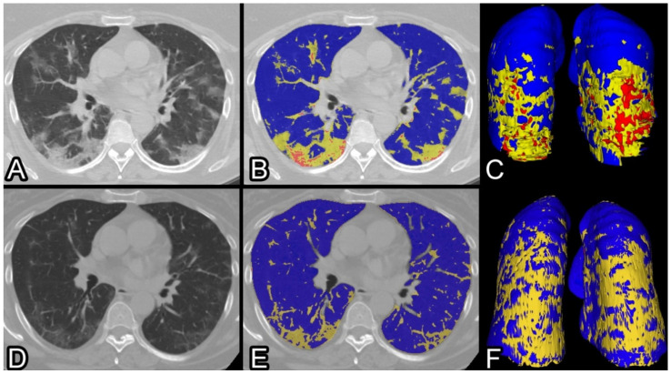
Figure 1.
CT scans of a 57 years old lady affected by COVID-19 pneumonia, treated with non-invasive oxygenation during 11 days of hospitalization. At admission, she lamented dyspnea at rest, cough, thoracalgia and fever. (A) Non-contrast CT scan ad admission showing typical bilateral ground-glass consolidation (B) QCT of the same scan highlighting in yellow the poorly aerated and in red the non-aerated lung volumes (C) three-dimensional view of the whole lungs. After 44 days, a CT scan shows significant improvement (D) with subtle residual ground-glass opacities and linear scarring, better outlined at QCT (E) and using 3D reconstruction (F). At the follow-up clinical assessment, the patient lamented persisting laboured breathing and abrupt intermittent coughing. Residual %CL and %PAL were 6% and 5%, consequently %deltaCL and −%deltaPAL −12% and −10%.

