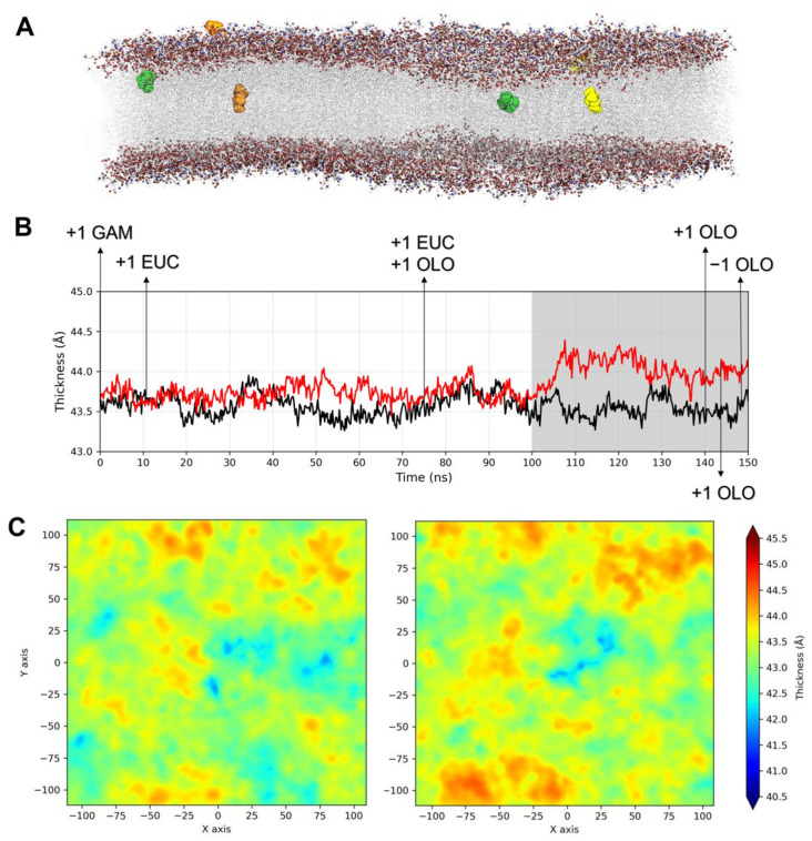
Figure 2.
(A) Binding locations observed for the γ-terpinene (orange), terpinen-4-ol (yellow), and 1,8-cineole (green) molecules, shown as spheres, within the viral membrane bilayer; (B) membrane thickness as a function of the simulation time in the absence (black line) or presence (red line) of the TTO compounds. Arrows indicate the time-points at which the entry (+1) or exit (−1) of the molecules was observed, and the grey background highlights the thickness shift; (C) thickness heatmaps for the viral membrane in the absence (left) or presence (right) of the TTO molecules.

