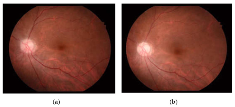
Figure 1.
Fundus photography of the left eye (a) on initial examination showed pinkish optic disc with optic disc edema (Frisen scale grade 2), and (b) after treatment with oral prednisolone, the optic disc edema resolved, and the optic disc became pale with a gliotic appearance in the temporal margin.

