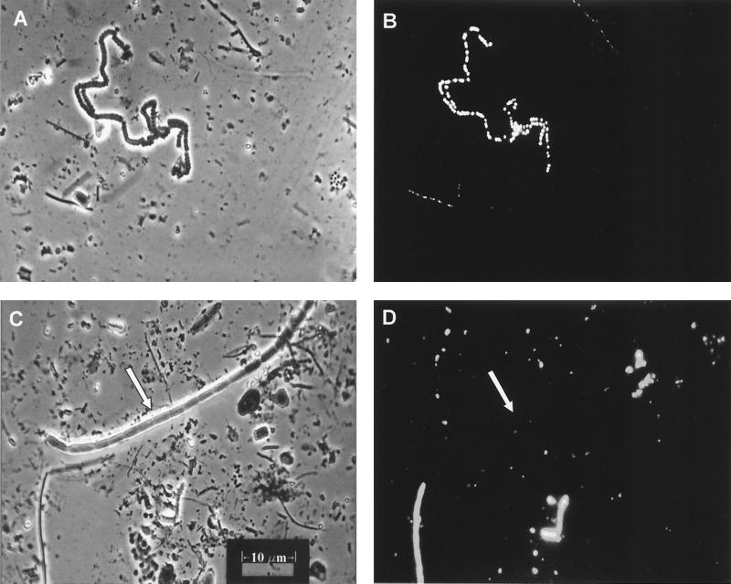
FIG. 7.
Micrographs of Nile Blue A-stained cells prepared directly from cyano/green layer mat material. (A) Exposure of a stained slide under incandescent light. (B) Same field as in panel A but exposed under fluorescent light. The fluorescing PHA granules of the cyanobacterium morphotype in the upper left of the field are distinctly visible. (C and D) Incandescent and fluorescent exposures, respectively, of another slide of the green layer material. Note the very large cyanobacterial filament marked by the arrow in panel C. This organism is not visible in panel D, indicating that it did not contain PHA granules.

