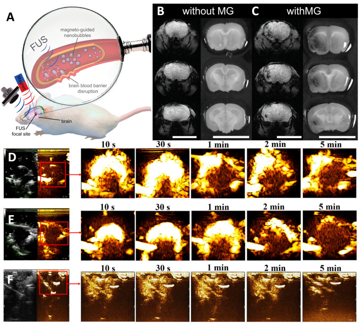
Figure 3.
(A) Nanobubbles can be used to diagnose locally disrupted BBB and accumulate due to magnetic guidance (MG). (B) Representative images of T2 gradient brain slices and their corresponding dye-treated brain tissue slices, used to assess the efficiency of bubble-free and magnetically guided brain tissue BBB disruption. (C) Biosafety induced by focused ultrasound compared to different degrees of BBB destruction caused by nanobubbles. Contrast-enhanced ultrasound imaging of brain tumors at other timepoints (10 s to 5 min) before and after injection of (D) nanobubbles and (E) commercial SonoVue. (F) Brain cavity ultrasound images at the same timepoint before and after the injection of NBs. Red squares show image enhancement of specific tissue sites. Adapted with permission from Refs. [103,104]. Copyright 2020 Elsevier and 2014 John Wiley and Sons.

