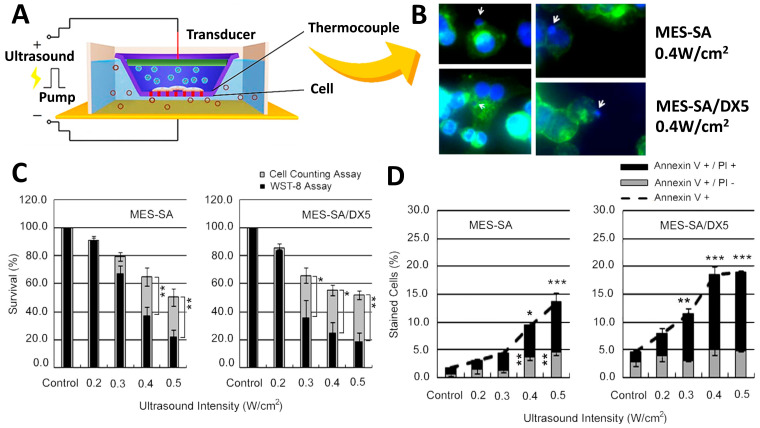
Figure 4.
(A) Schematic of the apparatus used to expose cells to ultrasound in vitro. Cells are cultured in transwells, and liposomes deliver siRNA or shRNA to inhibit the drug resistance of cancer cells. (B) After sonication at 0.4 W/cm2 for 24 h, the differences and sensitivity results of drug-sensitive uterine sarcoma cell lines before and after adding lipospheres (DX5) were observed with conjugate focus microscopy. White arrows indicate nuclear budding and demonstrate the inhibition of cells after adding lipospheres. Cell viability was assessed by: (C) WST-8 and cell-counting assays, showing that, after adding DX5 lipospheres, the viability of the cells was significantly inhibited. (D) Flow cytometric analysis of FITC-labeled Annexin V showing the cytostatic conditions. Asterisks (*) indicate the statistical significance of the difference between the absolute percentages obtained from cell counting assays, * p < 0.05, ** p < 0.1, and *** p < 0.01 considered significant. Adapted with permission from Ref. [107]. Copyright 2012 PLOS.

