Figure 7.
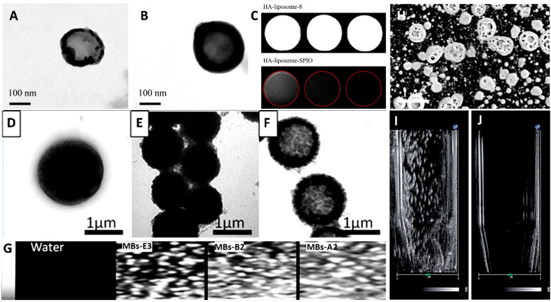
Different types of lipid spheres are used as contrast agents for ultrasound imaging. (A) Hydroxyapatite-coated liposomes and (B) hydroxyapatite-coated liposomes with superparamagnetic iron oxide. (C) MRI analysis with different liposomes. Microbubbles were modified with (D) polyamine salt, (E) magnetic polyamine salt, and (F) Fe3O4 nanoparticles. (G) Ultrasonic contrast images with microbubbles. (H) SEM images of nanobubbles. The ultrasonic wave was treated to a break of nanobubbles for (I) 0 s to (J) 5 min. Adapted with permission from Refs. [111,112]. Copyright 2011 and 2016 Elsevier and 2018 The Royal Society of Chemistry.

