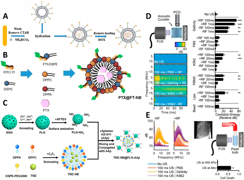
Figure 8.
Schematic illustrations showing the structure and biological functions of the (A) bubble-generating liposomes loaded with doxorubicin, (B) nanobubble-embed PTX, and (C) inorganic/nanobubble-conjugated nanocomposites with temozolomide loading. (D) Schematic of a passive cavitation detection setup using a 10 MHz transducer quadrature positioned to a focused ultrasound transducer. The broadband signal of cavitation was demonstrated with the Definity positive control. However, there was no cavitation in the PBS, and the only reflector cavitation was present in the K562 suspension. The group with only air bubbles showed a positive correlation between the cavitation energy and the cell destruction fraction, and exhibited significant thermal cavitation. (E) Waveforms in the K562 sample were induced by a 100 ms ultrasonic transducer with a reflector to form cavitation bubbles. The pipette bulb was pressurized to 400 kPa to create a pressure chamber. Under the overpressure of thermal cavitation, cell division was inhibited. Significant cavitation (compared to “No US”, ** p < 0.01, and *** p < 0.001) observed with definity. Adapted with permission from Refs. [114,117,118,119]. Copyright 2020 Future Medicine Ltd., 2017 Elsevier, 2021 American Chemical Society, and 2020 AIP Publishing.

