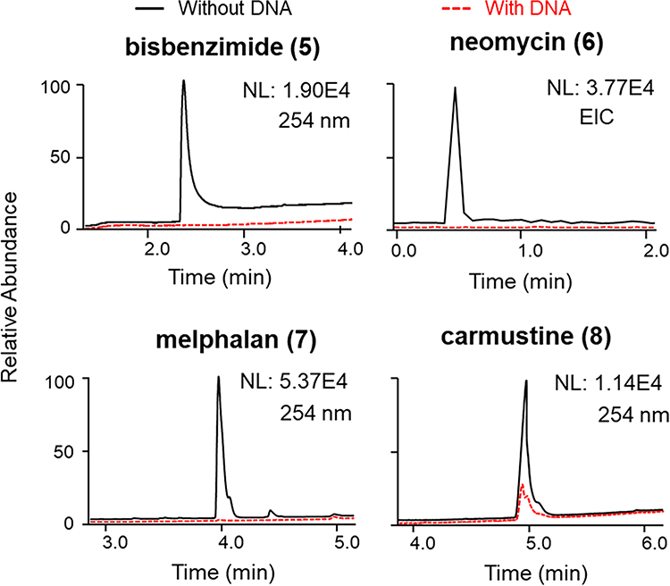
Figure 3.
Detection of groove-binding agents bisbenzimide (5) and neomycin (6) and covalent-binding compounds melphalan (7) and carmustine (8) using LLAMAS. Compounds 5, 7, and 8 were observed by UV (λ 254 nm) detection, whereas 6, which lacks a suitable UV chromophore, was monitored using the MS EIC trace. Individual plots show the peak areas for the compounds in the filtrate of the control group (incubated without DNA) superimposed on the traces recorded for the experimental samples (incubated with DNA) (NL: normalized intensity; EIC: extracted-ion chromatogram).

