
Fig. 6.
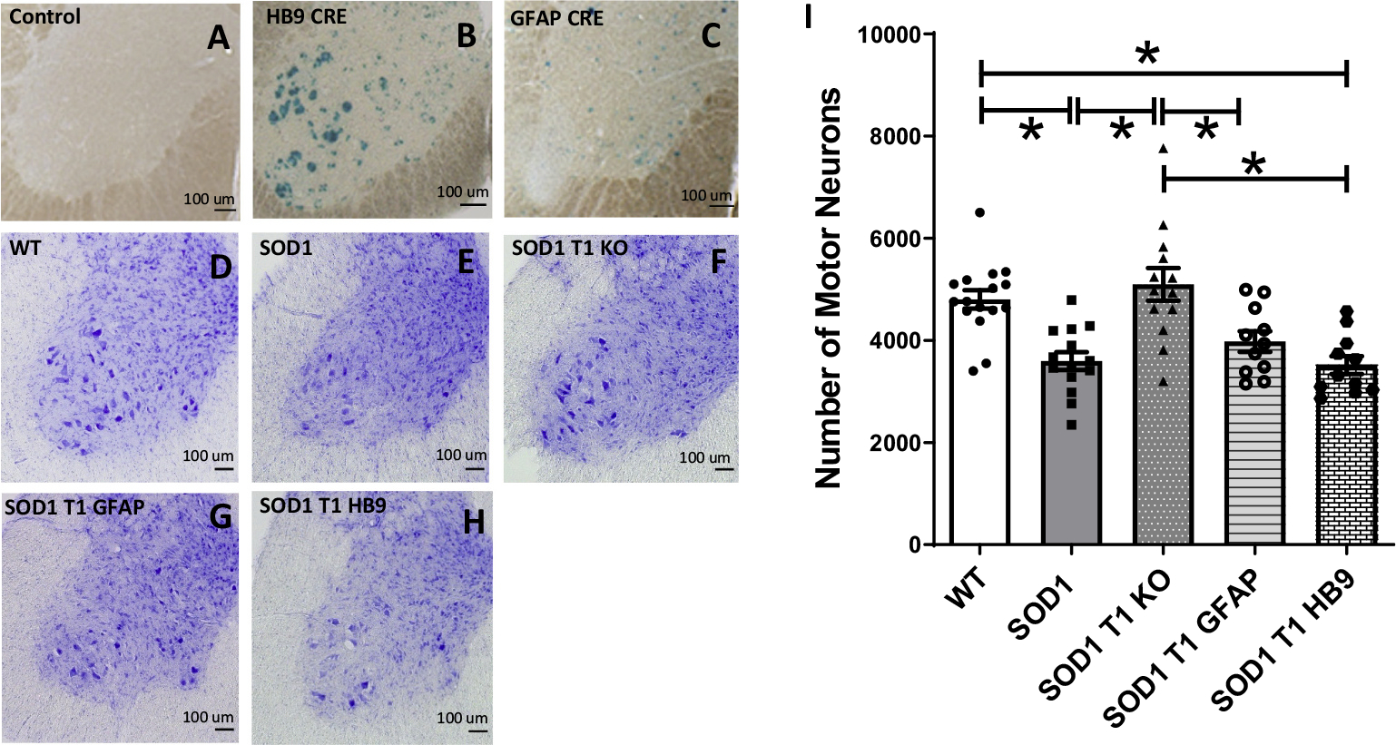
Deletion of TrkB.T1 in motoneurons or astrocytes of SOD1 mutant mice does not rescue motoneuron death at 12 weeks of age. (A-C) Representative β-Galactosidase staining images of lumbar spinal cords from a ROSA26-LacZ reporter mouse, used as negative control, (A), or a ROSA26-LacZ mouse crossed to a HB9-cre or a GFAP-cre transgenic showing specific cre activity in motor neurons (B) and astrocytes (C) respectively. (D—H) Representative Nissl staining of lumbar spinal cord sections of 12-week-old WT (D), SOD1 mutant (E), SOD1-TrkB.T1 KO (F), and SOD1 mice with an astrocytes (G; SOD1 T1 GFAP) or motoneuron-specific (H; SOD1 T1 HB9) TrkB.T1 knockout. (I) Histogram showing quantification of motor neuron numbers in the different mouse groups. Note that contrary to the SOD1 mutants with complete TrkB.T1 knockout showing rescue of motoneurons at 12 weeks, SOD1 mutants with specific TrkB.T1 deletion in either motoneurons or astrocytes are indistinguishable from SOD1 transgenic mice. Analysis of data was done by ANOVA followed by post-hoc Tukey’s multiple comparison test. * indicates p < 0.05. N = 11–16/group (WT, n = 16; SOD1, n = 14; SOD1 T1, n = 12; SOD1T1 GFAP, n = 11; SOD1T1 HB9, n = 11).