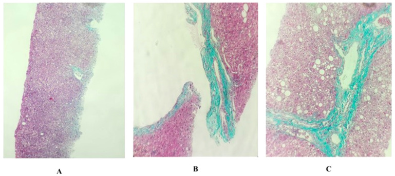
Figure 1.
Histopathology of liver tissues under 10× microscope. The METAVIR fibrosis staging system (F0, F1, F2): (A) F0 (the core needle biopsy shows liver tissue with two central veins, including the infiltration of some chronic inflammatory cells, many hepatocytes with hydropic degeneration and a few hepatocytes with fatty degeneration, but there is no appearance of portal zones); (B) F1 (the core needle biopsy shows liver tissue with one portal zone and no central veins, including the infiltration of numerous chronic inflammatory cells, a strong development of connective tissue surround portal zone, the bile duct is expanded and many hepatocytes contain fatty degeneration, but there is no appearance of bridging); (C) F2 (the core needle biopsy shows liver tissue with one or two portal zones and probably one central vein, including the infiltration of numerous chronic inflammatory cells, a strong development of connective tissue surrounding portal zones, many bile ducts are expanded, many hepatocytes contain large fatty degeneration and there is probably the appearance of bridging between central vein and portal zone).

