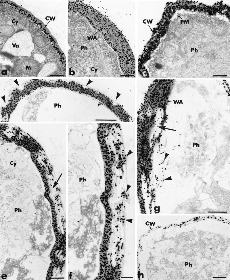
FIG. 7.
Transmission electron micrographs of mycelial samples collected from agar-coated slides 2 to 6 h after inoculation of P. parasitica (Ph): cellulolytic effect of P. oligandrum (Po) culture supernatant on P. parasitica hyphae. (a) P. parasitica exposed to PDB (control). Hyphae are delimited by a regularly labeled cell wall (CW) and contain a dense cytoplasm (Cy) in which mitochondria (M) and vacuoles (Va) are visible. Bar, 0.25 μm. (b) After 2 h of exposure to the culture supernatant of P. oligandrum, local retraction of the plasma membrane is accompanied by formation of wall appositions (WA). Bar, 0.5 μm. (c) After 3 h of exposure, complete retraction of the plasma membrane (PM), condensation of the cytoplasm, and loss of cell wall rigidity are detected. Bar, 0.25 μm. (d through g) Upon prolonged exposure (4 h or more), a large number of unlabeled areas are detected in the outermost wall layers of Phytophthora hyphae (panel d, arrowheads). These areas can extend to large portions of the cell wall (panel e, arrow) in which labeling is restricted to small wall fragments (panel f, arrowheads). Alteration of the wall appositions (panel g, large arrow) leads to release of labeled fragments (panel g, arrowheads). (d) Bar, 1 μm; (e) bar, 0.5 μm; (f) bar, 0.25 μm; (g) bar, 0.5 μm. (h) After 6 h of exposure, Phytophthora hyphae are surrounded by a slightly labeled cell wall. Bar, 0.5 μm.

