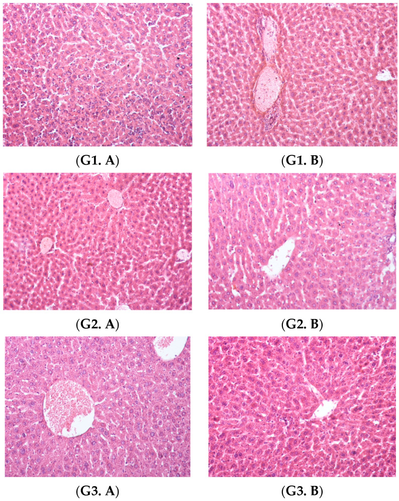
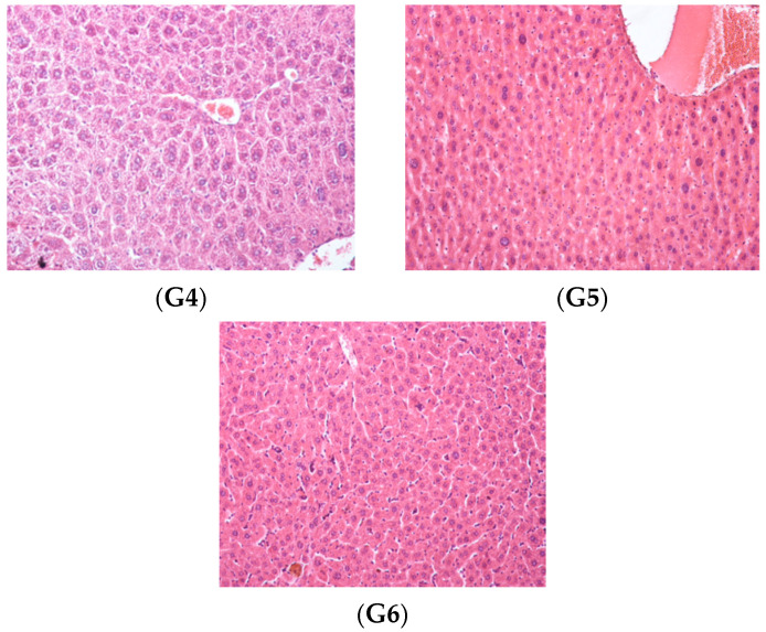
Figure 8.
Liver, hematoxylin and eosin staining × 20: Group 1, (G1. A) sinusoidal dilatations, capillary mononuclear cell infiltrations and the onset of necrosis and (G1. B) small areas of perivascular inflammation; Group 2, (G2. A) areas of vascular congestion and (G2. B) sinusoidal dilatations and reduced areas of inflammation; Group 3, (G3. A) extensive vascular congestion and (G3. B) slight sinusoidal dilatations; Group 4 (G4), hepatocellular vacuolation and reduced vascular congestion; Group 5 (G5), normal liver morphology; Group 6 (G6), normal liver morphology.


