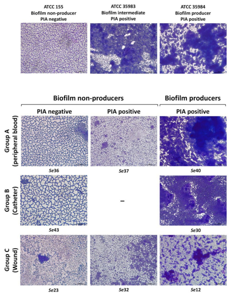
Figure 5.
The visualization of the biofilm structure and matrix production among the S. epidermidis clinical isolates. Giemsa staining of the biofilms of the S. epidermidis reference strains (upper panel) and clinical isolates (lower panel) showing the different biofilm architectures formed by bacteria cultured on glass coverslips. The name of each clinical strain, subdivided according to their biofilm and PIA production, has been reported. Images were taken with a 200× magnification, the scale bars (bottom right corner of each image) represent 100 μm.

