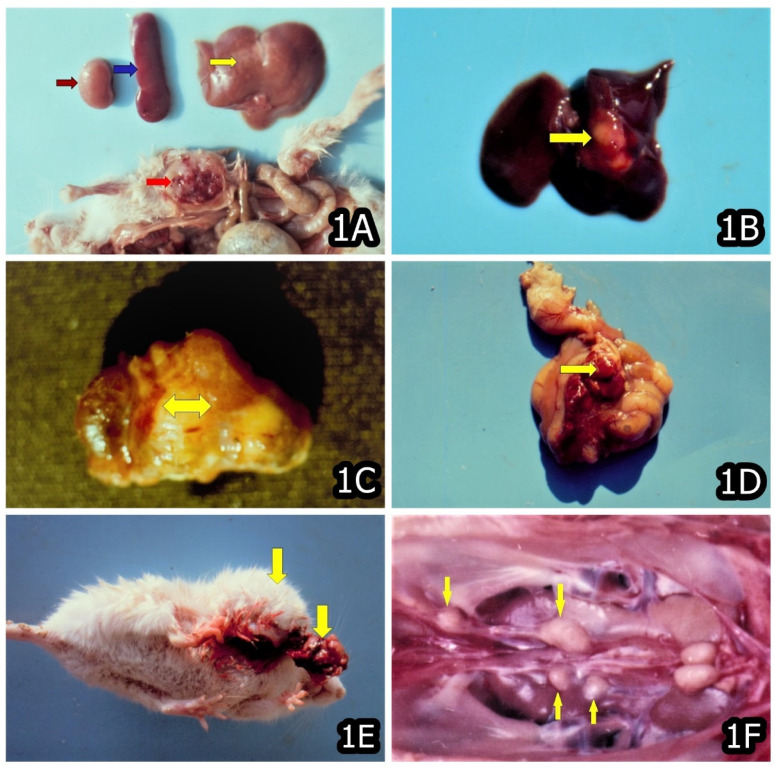
Figure 1.
(A) Rhabdomyosarcoma in the muscles (red arrow), necroses in the liver (yellow arrow) and enlargement of the spleen (blue arrow) and pale color of kidney (brown arrow) in a mouse that died between months 15–20 and was exposed to 10 ppm OTA and 50–60 ppm PA. (B) Carcinoma in the liver (yellow arrow) in a mouse that died between months 10–15 and was exposed to 10 ppm OTA and 50–60 ppm PA. (C) Carcinoma in the region of kidney (yellow arrow) in a mouse that died between months 15–20 and was exposed to 10 ppm OTA and 50–60 ppm PA. (D) Angiosarcoma in the intestinal mesenterium (yellow arrow) in a mouse that died between months 15–20 and was exposed to 10 ppm OTA and 50–60 ppm PA. (E) Subcutaneous sarcoma (yellow arrows) in a mouse that died between months 15–20 and was exposed to 10 ppm OTA. (F) Carcinoma in the region of ureters (yellow arrows) of male chick exposed to 5 ppm OTA via the feed, which died at the end of the 20th month of the experiment. Large grey-white neoplastic foci are seen along the ureters and protruded significantly above its surface.

