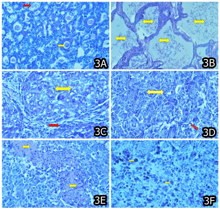
Figure 3.
(A) Adenocarcinoma in the kidney containing secreted substance or hyaline (yellow arrow) and and/or a few necrotic tumor cells (red arrow) in a mouse at the end of 20 months exposure to 10 ppm OTA and 50–60 ppm PA. (B) Adenocarcinoma lined with several layers of neoplastic cells and containing a secreted substance and/or necrotic tumor cells and leucocytes (yellow arrow) in the liver in a mouse that died between months 15–20 and was exposed to 10 ppm OTA and 50–60 ppm PA. (C) Adenocarcinoma (nest adenocarcinoma—yellow arrow) in the liver with well-formed nests of malignant tumor cells surrounded by a relatively well-developed stromal tissue (red arrow) in a mouse that died between months 10–15 and was exposed to 10 ppm OTA and 50–60 ppm PA. (D) Carcinoma solidum (yellow arrow) with scarce stromal tissue (red arrow) in the liver of a mouse that died between months 10–15 and was exposed to 10 ppm OTA and 50–60 ppm PA. (E) Carcinoma solidum with many necroses (yellow arrow) among the neoplastic tissue infiltrated with leucocytes in the liver of a mouse that died between months 10–15 and was exposed to 10 ppm OTA and 50–60 ppm PA. (F) Sarcoma mixtocellulare consisted of globular cells with different size, polymorphism, polychromasia (yellow arrow), low differentiation, many irregular mitoses of the cells and their nuclei in the subcutaneous tissue in a mouse that died between months 15–20 and was exposed to 10 ppm OTA.

