Figure 4.
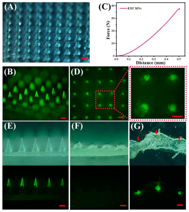
Characterization and insertion capability of TS-MNs. (A) Stereomicroscopic images of TS-MNs containing FITC-EXT. Scale bars, 200 μm. (B) Fluorescence images of TS-MNs encapsulated FITC-EXT. Scale bars, 200 μm. (C) Mechanical behavior of the TS-MNs. (D) Fluorescence images of porcine skin after TS-MNs application and removal. Scale bars, 200 μm. Side view of stereomicroscope (above) and fluorescence microscopy (below) of TS-MNs before (E) and after (F) skin insertion for 30 min. Scale bars, 200 μm. (G) Histological section of isolated porcine skin imaged by stereomicroscope (above) and fluorescence microscopy (below). Scale bars, 200 μm.

