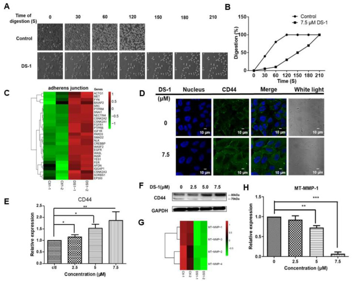
Figure 7.
DS−1 enhanced U−2 OS cells’ adhesion. (A) The morphological changes of U−2 OS cells treated with/without DS−1. Cells were treated with 0 and 7.5 μM DS−1 for 24 h, washed with PBS, and digested with trypsin at 25 °C under a microscope, and photos were taken every 30 s. The percentages of digested cells are recorded in (B). (C) Heatmap of genes related to the adherens’ junction in the control group and DS−1 group. (D) U−2 OS cells were treated with 0 and 7.5 μM DS−1 for 24 h, and the expression of CD44 was observed by immunofluorescence. (E,F) CD44 and GAPDH were analyzed by Western blot and calculated by Image J. (G) The relative expression of 4 kinds of MT-MMPs was analyzed by the transcriptome, as shown in the heatmap. (H) After being treated with DS−1, the relative expression of MT-MMP-1 was verified by RT-PCR. Values are the average of three independent experiments, * p < 0.05, ** p < 0.01, *** p < 0.005.

