Figure 8.
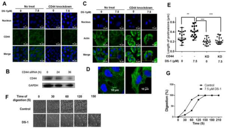
CD44 knockdown reduced cell adhesion by shortening cell protrusions. (A) U−2 OS cells transfected with CD44 siRNA and no siRNA were seeded into a 24-well plate treated with 7.5 μM DS−1 for 24 h, and the expression of CD44 was detected by immunofluorescence. (B) CD44 expression of U−2 OS cells was detected by Western blotting after being treated with siRNA for 0, 24, and 36 h. (C) U−2 OS cells were first transfected with/without CD44 siRNA and then treated with DS−1 for 24 h, and its actin morphologies were detected. The representative magnified actin morphologies are listed in (D). (E) The lengths of protrusions in (C) were analyzed by Image J and are shown in a scatterplot graph. The lengths of 20 protrusions from actin in (C) were calculated. Each value is the average of 20 lengths of protrusions, ** p < 0.01, *** p < 0.005. (F) U−2 OS cells were transfected with CD44 siRNA, then treated with 0 and 7.5 μM DS−1 for 24 h and digested with trypsin at 25 °C; the digestion time was recorded by a microscope, and photos were taken every 30 s. The percentage of digested cells is recorded in (G).

