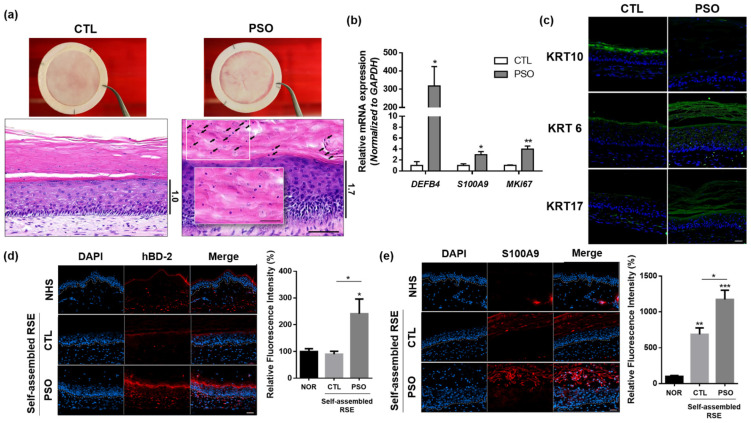
Figure 4.
Psoriatic phenotypes of the IL-17A-induced self-assembled RSE models. (a) histological analysis of self-assembled psoriasis RSE model (Bar = 100 µm, n = 4). The arrows indicate remaining nuclei in the stratum corneum. (b) The mRNA expression of DEFB4, S100A9, and MKi67 in the self-assembled RSE model was analyzed by RT-qPCR. (c) Immunofluorescence staining analysis of KRT6, KRT10, and KRT17 in self-assembled psoriasis RSE model (Bar = 50 µm, n = 4). (d) Immunofluorescence staining of HBD-2 in normal human skin and self-assembled RSE (Bar = 50 µm, n = 4). For quantification, the fluorescence intensity of the red signals was examined by Image J. (e) Immunofluorescence staining of HBD-2 in normal human skin and self-assembled RSE (Bar = 50 µm, n = 4). For quantification, the fluorescence intensity of the red signals was examined by Image J. The self-assembled RSE models were treated with vehicle (D.W) for control or 100 ng/mL of IL-17A for the psoriatic model is treated to the culture medium. RSE: Reconstructed skin equivalent; NHS: Normal human skin; PSO: Psoriatic RSE. * p < 0.05; ** p < 0.01; *** p < 0.005.

