Figure 1.

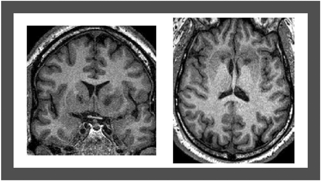
T1-weighted magnetic resonance imaging acquired 12 months post-surgery showing double shot GVC bilateral lesions of the ventral portion of the internal capsule, visualized in the coronal (left) and axial (right) planes.

T1-weighted magnetic resonance imaging acquired 12 months post-surgery showing double shot GVC bilateral lesions of the ventral portion of the internal capsule, visualized in the coronal (left) and axial (right) planes.