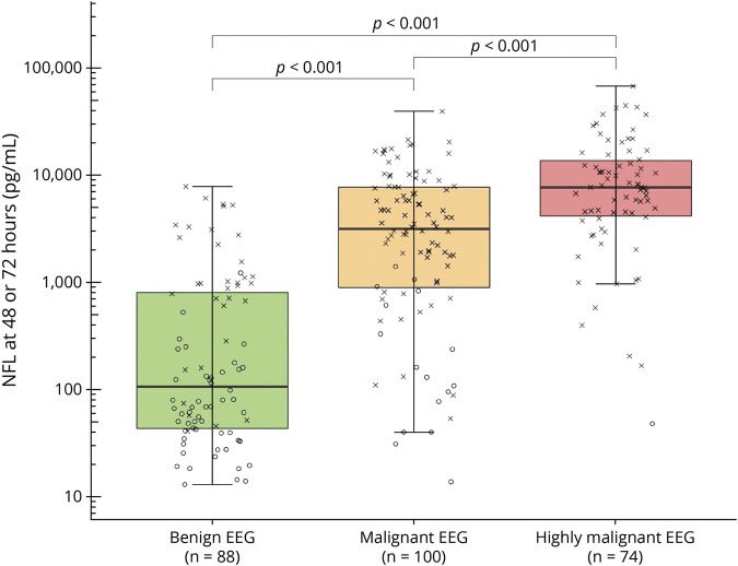
Figure 2. Highly Malignant, Malignant, and Benign EEG Patterns and Serum NfL.
Boxplot demonstrating logarithmic peak neurofilament light (NfL) levels (highest serum neurofilament levels at either 48 or 72 hours postarrest) for EEG patterns as defined by Westhall et al.7: “highly malignant”: burst-suppression or suppression with or without discharges; “malignant”: discontinuous, reversed anterio-posterior gradient or low-voltage background, abundant rhythmic or periodic discharges or unequivocal seizures; “benign”: continuous background without malignant features. Neurologic outcome for each patient is indicated through “X” (poor outcome, Cerebral Performance Category [CPC] 3–5) or “O” (good outcome, CPC 1–2) at 6 months’ follow-up. Peak NfL was increasingly higher in more malignant EEG patterns (p < 0.001).

