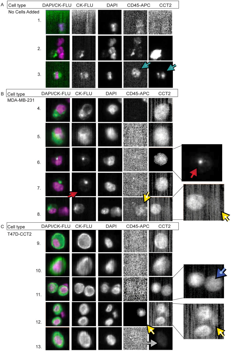
Fig 4. Breast cancer cells spiked in blood can be detected based on CCT2 staining using the CSS.
Representative images of (A) whole healthy human blood without spiked cancer cells processed through the CSS and stained for CCT2, and (B-C) Representative images of MDA-MB-231 (B) or T47D-CCT2 (C) cells spiked into human blood, processed through the CSS, and stained for CCT2. Light blue arrows: leukocyte that is CCT2 positive. Red arrows: cells with dim CK signal and CCT2 positive signal. Yellow arrows: leukocytes that have dim CCT2 signal. Dark blue arrow: doublet of spiked cancer cells with different CCT2 staining intensities. Grey arrows: cells with dim CCT2 signal. CK-FLU. This data is representative of ten experiments.

