Fig. 3.
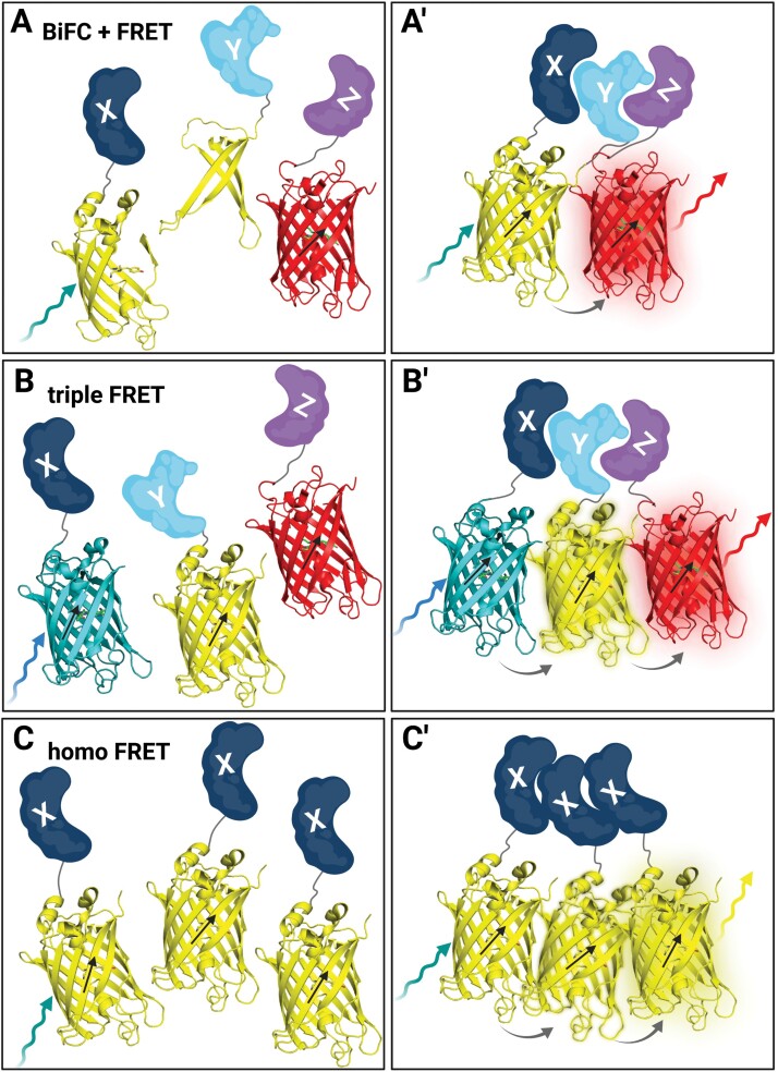
Techniques to measure PPI of more than two POIs. (A) In a combined BiFC–FRET experiment, two POIs (X, dark blue; Y, light blue) are fused to the N- or C-terminal part of a split FP (here, split 3D YFP structures in yellow), and a third POI (Z, purple) is fused to an acceptor FP (here, 3D mCherry structure in red). (Aʹ) In the case of interaction of all three POIs, the YFP parts are reconstituted and, after excitation (teal arrow), can transfer energy by FRET (grey arrow) to the acceptor FP (mCherry) which can emit light (red arrow). (B) In a triple FRET experiment, three POIs (X, dark blue; Y, light blue; Z, purple) are fused to three different FPs (here, 3D structures of CFP, cyan; YFP, yellow; and mCherry, red). (Bʹ) In the case of interaction of all three POIs, the three FPs come close enough to allow, upon excitation of CFP by blue light (blue arrow), energy transfer (grey arrow) by FRET to YFP as acceptor which then serves also as a donor and transfers energy via FRET (grey arrow) to mCherry which can then emit light (red arrow). (C) In a homo-FRET experiment, two or more of the same POI (X, dark blue) are labelled with an FP (here, 3D structure of YFP in yellow). (Cʹ) If the POI can form higher order complexes, energy can be transferred from one excited (teal arrow) FP to another, thereby depolarizing the emission of the FP (yellow arrow). Figure created with BioRender.com.

