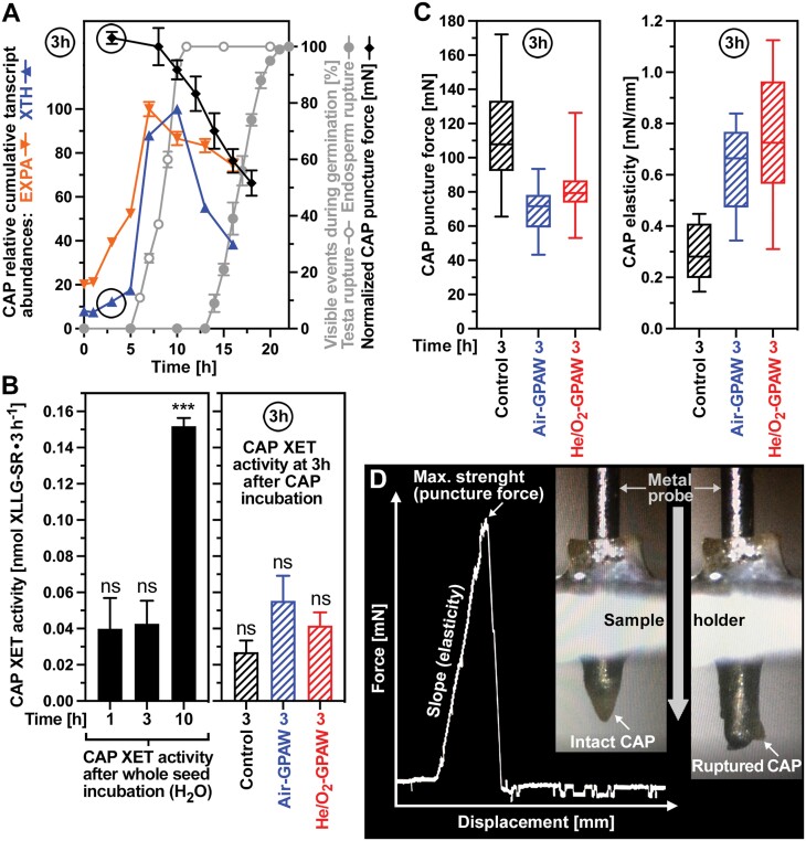
Fig. 5.
Biomechanical and biochemical analysis of GPAW-induced endosperm weakening. (A) Time courses of micropylar endosperm (cap) puncture force, testa and endosperm rupture, and expansin (EXPA) and xyloglucan endotransglycolases/hydrolase (XTH) transcript abundances in the cap of Lepidium sativum FR14 seeds during germination. For the cap puncture force, normalized values combined from two datasets (Linkies et al., 2009; Graeber et al., 2014) are presented. The cap-specific relative cumulative expression values of EXPA and XTH genes were from the spatiotemporal transcriptome dataset of L. sativum seed germination (Scheler et al., 2015); for individual genes and details, see Supplementary Fig. S4. (B) Xyloglucan endotransglycosylase (XET) enzyme activities of XTH proteins in the cap after incubation of whole L. sativum seeds in dH2O for the times indicated (left panel). Effects of incubating isolated caps in Air-GPAW (45 min) or He/O2-GPAW (45 min) on the XET enzyme activities at 3 h (right panel). Note that only the 10 h CAP XET activity was statistically different, while all of the other XET activity values were not significantly (‘ns’) different from each other. (C) Biomechanical analysis of the effects of Air-GPAW (45 min) or He/O2-GPAW (45 min) on cap endosperm weakening at 3 h. The cap puncture force (tissue resistance) at 3 h (left panel) was determined as the maximal force from the displacement–force curve, and the cap tissue elasticity at 3 h (right panel) was calculated as the slope of the linear portion of the displacement–force curve. (D) Example displacement–force curve and images of the biomechancial assay with cap prior to rupture (intact) and cap post-rupture by the metal probe of the biomechanics device.

