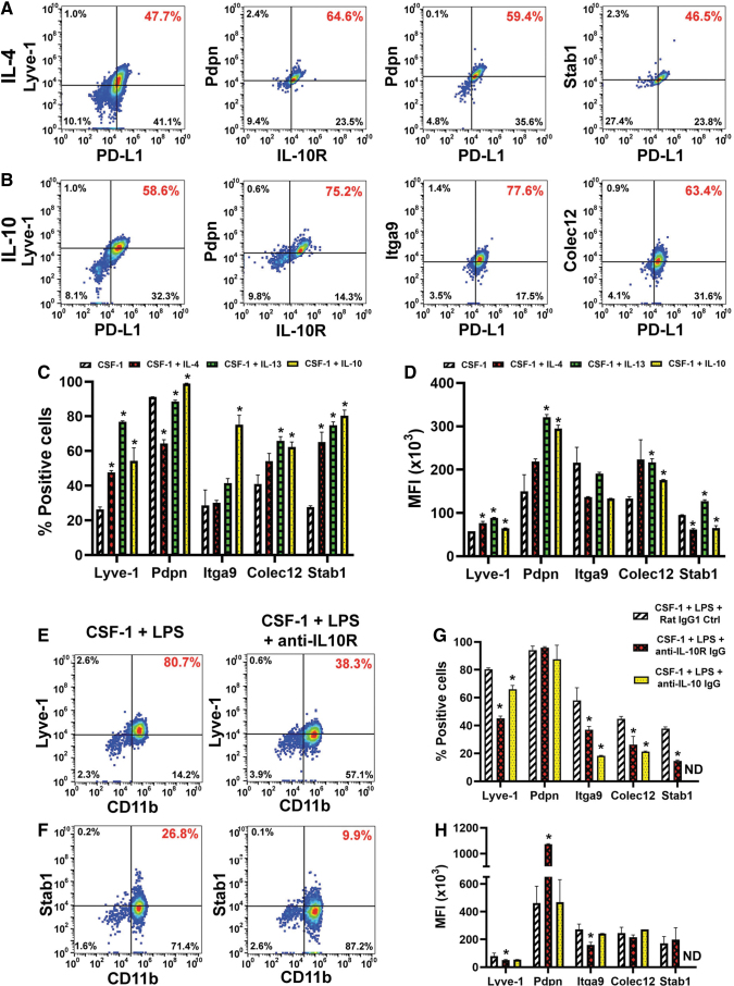
FIG. 4.
Th2 cytokines promote the lymphatic identity in CSF-1-primed myeloid precursors. BM cells were differentiated with CSF-1 and either (A) IL-4 or (B) IL-10. Flow cytometry dot plots of cells stained for M2 markers PD-L1 or IL-10R and lymphatic specific proteins Lyve-1, podoplanin (Pdpn), integrin-a9 (Itga9), collectin-12 (Colec12), or stabilin-1 (Stab1). Percentage of double-positive cells is highlighted in red font. (C) The mean percent of positive cells and (D) MFI ( × 103) ± SD for each LEC target. Statistical significance between cells differentiated with Th2 cytokines and CSF-1 alone determined by a Student's t-test with P values ≤0.05 and it is indicated by *. (E–H) Standard differentiation of BM cells by CSF-1/LPS was performed in the presence of control or blocking antibodies to IL-10R or IL-10 ligand. On day 6, cells were stained for CD11b in combination with anti-Lyve-1 (E) or anti-stabilin-1 (F) antibodies. Percent of double-positive cells for each dot plot is identified in red font. (G) The mean percent of positive cells ± SD and (H) mean MFI ( × 103) ± SD for each LEC marker expressed in control and IL-10 pathway blocking antibodies. Statistical significance between marker expression in differentiated cells in the presence of control and blocking antibodies was determined by a Student's t-test with P values ≤0.05 and it is indicated by *. ND (not done) indicates markers that were not analyzed in some assays. Each assay was performed in duplicate and reproduced in three independent experiments. LEC, lymphatic endothelial cell.

