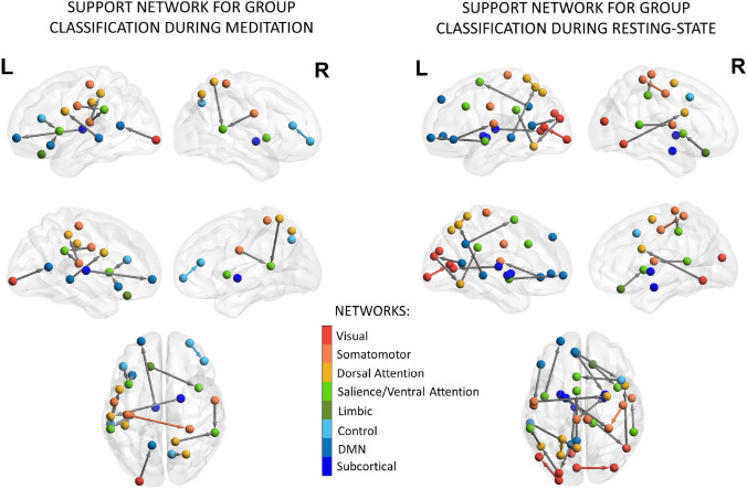
Fig. 5.
RFE results: support network for group classification during meditation. The group signatures (i.e., highest-ranked EC features) are presented for the two conditions in sagittal, medial, and dorsal views, both for the right and left hemispheres. The color of the nodes is determined by their belongingness to one of the eight resting-state networks (red = visual; orange = somatomotor; light orange = dorsal attention; light green = salience/ventral attention; dark green = limbic; light blue = control; blue = DMN; dark blue = subcortical). The networks are drawn from the parcellation in 100 ROIs comprising 7 functional divisions (Schaefer et al. 2018) together with 16 subcortical regions (Tian et al. 2020). Gray arrows show the directionality of connection (EC-links) between ROIs of distinct networks. In contrast, direct links between nodes belonging to the same network are colored according to network-specific hue. Visualization of results was generated using the BrainNet Viewer Toolbox (Xia et al. 2013)

