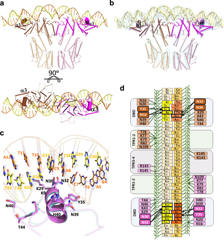
Fig. 5. AimRKat DNA-binding characterisation.
a Overall structure of AimRKat in complex with DNA. Two orthogonal views are shown. Subunits A and B are represented in cartoon with helices as cylinders and coloured in brown and magenta respectively. DNA is painted in orange and yellow for 5′-3′and 3′-5′ strands, respectively. Darker colours have been used for DNA interacting regions with helix α3 shown with a bigger radius and DNA interacting residues represented in stick. b Overall superposition of AimRKat (same colour code as for a) and AimRSPβ (coloured in blue). DNA is shown as yellow cartoon and surface for AimRKat and blue surface for AimRSPβ. c Detail view of DNA specific recognition mediated by helix α3 in AimRKat (coloured in magenta) and AimRSPβ (semi-transparent and coloured in blue). Interacting residues are shown in sticks and labelled. d Schematic representation of the DNA-AimRKat contacts. The colour code is the same as for (a). Sequence-specific interactions for DNA readout are highlighted with thicker lines and the residues carrying out these interactions with more intense colours.

