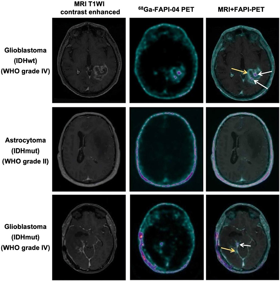
Fig. 2.
Axial view images (contrast-enhanced T1-weighted MRI, 68 Ga-FAPI-04 PET and fused images of both modalities) of an IDH-wildtype glioblastoma, an IDH-mutant astrocytoma, and an IDH-mutant glioblastoma. The IDH-wildtype glioblastoma and the high-grade IDH-mutant glioblastoma both show foci with moderately to markedly increased tracer uptake (white arrows and yellow arrows) within the contrast-enhancing lesion. In contrast, the tracer uptake showed only slightly elevated in IDH-mutant astrocytoma and very low in healthy brain parenchyma. Figure adapted from Rohrich et al. [46]

