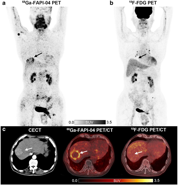
Fig. 5.
A 78-year-old man with moderately differentiated hepatocellular carcinoma (HCC) and liver cirrhosis. 68 Ga-FAPI-04 PET/CT (a, c) demonstrates an increased focal uptake (SUVmax = 4.05 and TBR = 3.93) in the liver, while 18F-FDG PET/CT (b, c) scans showed non-FDG-avid lesion (SUVmax = 2.55 and TBR = 1.20) indicated by arrows. Figure adapted from Guo et al. [64]

