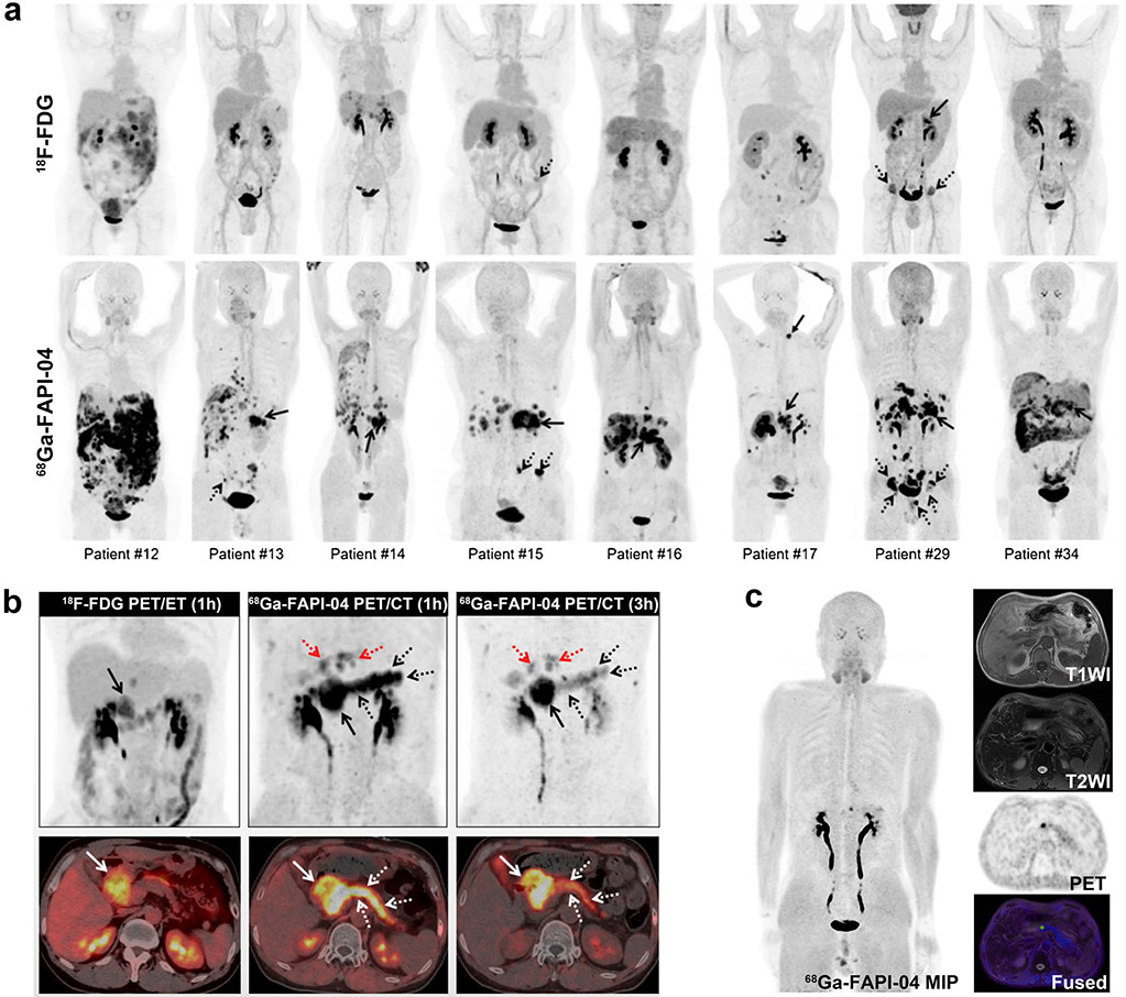
Fig. 6.
Eight patients with pancreatic cancer underwent 18F-FDG and 68 Ga-FAPI-04 PET/CT imaging. 68 Ga-FAPI-04 PET/CT outperformed 18F-FDG in detecting primary tumors (solid arrows), supraclavicular lymph node metastases (arrowhead), abdomen lymph node metastases, liver metastases, peritoneal carcinomatosis, and bone metastases (dotted arrows) (a). The patient with pancreatic cancer was visible the high metabolic activity of the primary tumor on the 18F-FDG PET/CT and 68 Ga-FAPI-04 PET/CT scan (solid arrows). However, the 1-h image for 68 Ga-FAPI-04 PET/CT (middle line) demonstrated intense 68 Ga-FAPI-04 uptake in the body and tail of the pancreas (white and black dotted arrows) and moderate FAPI uptake in the intrahepatic bile duct (red dotted arrows). 3-h delayed 68 Ga-FAPI-04 PET was performed to differentiate between malignant and benign lesions (right line). The SUVmax value of the primary tumor was slightly elevated, whereas the SUVmax values of the pancreatitis lesions were decreased compared to the 1-h standard image (b). A 53-year-old man suffered from retrosternal and laryngeal discomfort for more than 9 months. Laboratory testing revealed a high CEA level (11.3 μg/L). 68 Ga-FAPI-04 PET/MR revealed focal uptake in the pancreatic neck with a negative corresponding MR signal. Furthermore, no abnormal lesions were found in subsequent DCE-MRI and EUS (c). Figure adapted from Pang et al. [71] and Zhang et al. [72]

