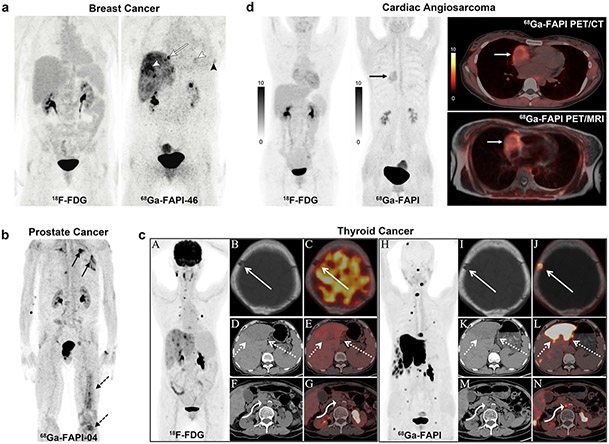
Fig. 7.
(a) A 60-year-old woman with triple-negative breast cancer. Neither liver and lung metastases (morphologically observed) nor local relapse demonstrated pathologic uptake in 18F-FDG PET. However, 68 Ga-FAPI-46 PET showed elevated tracer uptake in metastatic lesions (white arrowheads) and local relapse (black arrowhead). Additional osseous metastasis that was previously not discernible is noted (arrow). (b) A 70-year-old patient was diagnosed with CRPC. 68 Ga-FAPI-04 PET/CT demonstrated bone (dotted arrows) and pulmonary metastases (solid arrows). (c) A 76-year-old woman previously underwent total thyroidectomy surgery to treat papillary thyroid carcinoma. The 68 Ga-FAPI PET/CT showed more and higher metabolic lesions than 18F-FDG (it was unclear which specific FAPI-based tracer was used in this study, likely 68 Ga-FAPI-04). 18F-FDG PET/CT showed destruction of the skull without 18F-FDG uptake (solid arrow), liver mass with moderate FDG uptake (dotted arrows), and enlarged retroperitoneal lymph node with mild FDG uptake (curved arrow). The axial of 68 Ga-FAPI PET/CT showed destruction of the skull with intensive FAPI uptake, liver mass with intensive FAPI uptake, and enlarged retroperitoneal lymph node with moderate FAPI uptake. (d) A 31-year-old woman presented with a history of chest pain. Ultrasonography showed massive pericardial effusion, and subsequent pericardiocentesis drained the bloody pericardial effusion. The patient underwent 18F-FDG to detect the primary tumor; however, no abnormal activity was observed. Follow-up with 68 Ga-FAPI PET/CT was performed to improve detection (it was unclear which specific FAPI-based tracer was used in this study, likely 68 Ga-FAPI-04). A focus with increased 68 Ga-FAPI uptake in the right chest was revealed (black arrow). The axial PET/CT views localized this abnormality in the right atrium (white arrow), with a target/myocardium ratio of 3.9. Figure adapted from Backhaus et al. [76], Kesch et al. [86], Wu et al. [91] and Zhao et al. [94]

