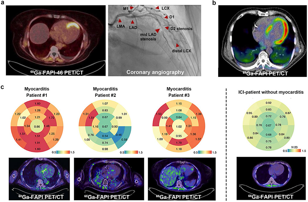
Fig. 9.
Representative 68 Ga-FAPI PET images of cardiovascular disease. (a) A 62-year-old woman presenting with STEMI underwent FAPI PET imaging 9 days after angiography. The subtotal stenosis of the mid-LAD is visualized perfectly matching the apical/apicoseptal tracer uptake in the PET scan. (b) A 67-year-old male patient with pancreatic ductal adenocarcinoma had received systemic cancer therapy with gemcitabine and Nab-Paclitaxel. During the disease, 68 Ga-FAPI PET showed an intensive tracer accumulation of the left ventricular myocardium, indicating it may be capable of detecting chemotherapy-induced myocardial injuries (it was unclear which specific FAPI-based tracer was used in this study, likely 68 Ga-FAPI-04). (c) 68 Ga-FAPI PET/CT illustrates ICI-associated myocarditis. Bulls Eye Illustration of SUVs showing their distribution in the myocardium of the left ventricle in 17 defined areas. The enrichment is shown for ICI-associated myocarditis patients #1-#3 (left panel). In comparison, the median signal of patients who have received immune checkpoint inhibitors without signs of myocarditis is summarized (right panel). STEMI, ST-segment elevation myocardial infarction; LAD, left anterior descending (artery); D1, D2, diagonal branches of LAD. Figure adapted from Kessler et al. [103], Totzeck et al. [110] and Finke et al. [113]

