
FIGURE 8.
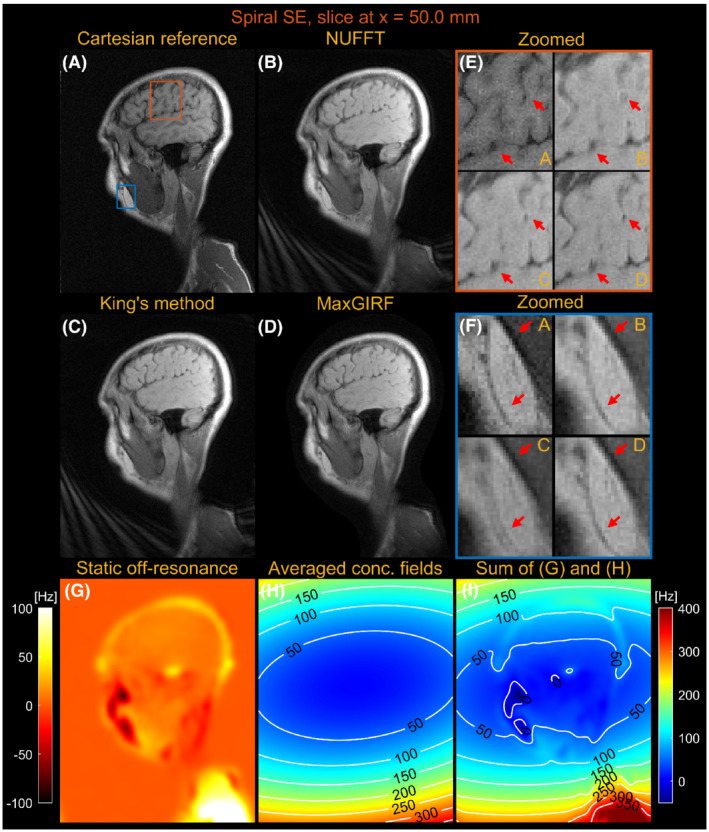
Sagittal spiral spin‐echo imaging of a healthy volunteer at 0.55 T off‐isocenter (x = 50.0 mm). Comparison of image reconstructions using comparator Cartesian spin‐echo image (A), NUFFT reconstruction (B), King's method without static off‐resonance correction (C), and MaxGIRF reconstruction with static off‐resonance correction (Low‐rank approximation L = 30) (D). E, Zoomed‐in image of a brain region (orange box). F, Zoomed‐in image (blue box). G, Static off‐resonance map. H, Time‐averaged concomitant fields map. I, Sum of the static off‐resonance map and time‐averaged concomitant fields map. King's method may adversely increase blurring artifacts (eg, blue box) compared with NUFFT reconstruction when the static off‐resonance in a region counteracts the concomitant fields. However, MaxGIRF with static off‐resonance correction correctly handles such regions as shown in (F) and provides “sharper” delineation of brain tissue boundaries in (E) compared with King's method