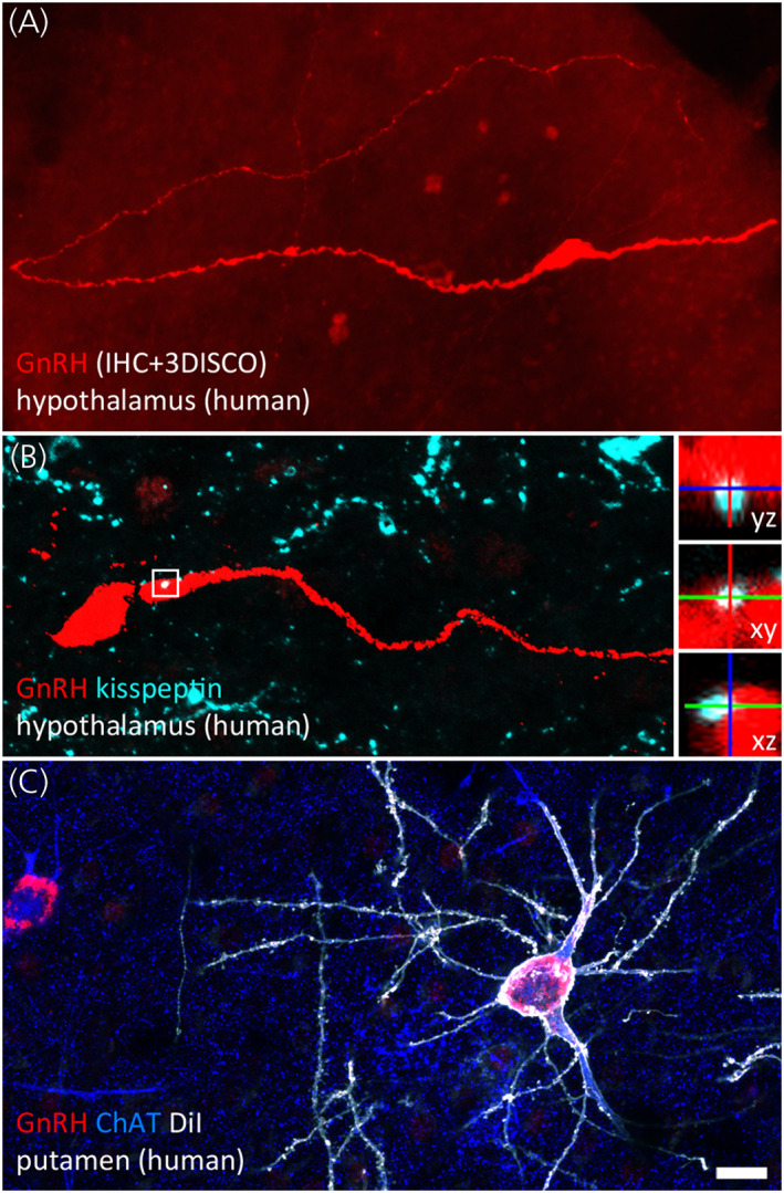
FIGURE 3.

Hypothalamic and extrahypothalamic GnRH neurons of the adult human brain. (A) Hypothalamic GnRH neurons regulating reproduction are typically fusiform. In 1 mm‐thick slices made transparent with the 3DISCO clearing technology, lengthy GnRH dendrites can be followed occasionally for several millimetres. Dendrites may represent the main cellular compartment receiving afferent inputs. (B) Kisspeptin‐immunoreactive inputs to GnRH neurons (turquoise) from the infundibular (arcuate) nucleus convey information about circulating sex steroid levels. High‐power insets show orthogonal views of a neuronal apposition. (C) Unlike rodents, humans contain hundreds of thousands of GnRH‐immunoreactive (red) neurons in extrahypothalamic brain regions. The dendritic tree of GnRH cells in the putamen can be visualized post mortem using the lipophilic dye DiI (shown in white) delivered to the sections with the aid of a Gene Gun. Labelled GnRH neurons exhibit smooth surfaced dendrites and correspond to a subpopulation of cholinergic interneurons immunoreactive to choline acetyltransferase (ChAT; blue). Scale bar: 20 μm in (A) and (C), 16 μm in (B) (insets: 5 μm). Photograph courtesy of Dr Katalin Skrapits, Institute of Experimental Medicine, Budapest
