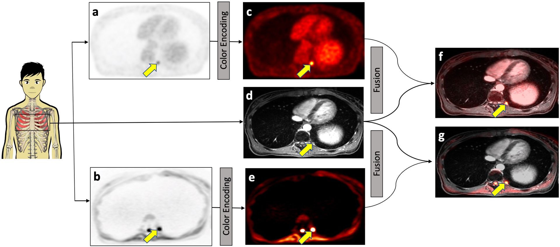
Fig. 2.

Concept of integrated 2-[18F]FDG-PET/MRI and DW-MRI. 2-[18F]FDG-PET (a) and DW-MRI (b) images were color-encoded (c and e, respectively) and then fused with the contrast-enhanced MRI scan (d) for anatomical orientation, yielding the integrated 2-[18F]FDG-PET/MRI (f) or DW-MRI (g). Both 2-[18F]FDG-PET and DW-MRI images visualize a subcentimeter bone marrow metastasis in the left proximal 11th rib (arrow) in a 25-year-old female patent with Wilms tumor.
