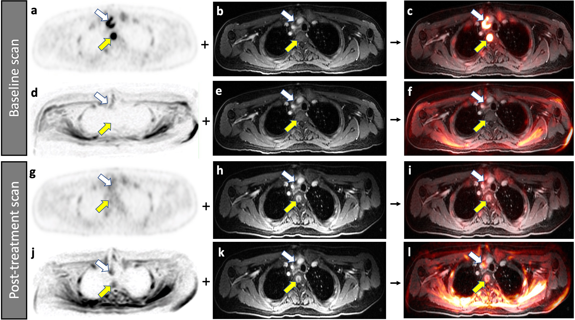
Fig. 4.

Improved detection of a bone marrow metastasis in a 22-year-old male patient with diffuse large B-cell lymphoma on 2-[18F]FDG-PET compared to DW-MRI. Baseline scans (a-f), A bone marrow metastasis in the third thoracic vertebra (yellow arrow) demonstrates high FDG uptake on 2-[18F]FDG-PET (a). The lesion is not visible on the contrast enhanced MRI (b). The combined 2-[18F]FDG-PET/MR scan localized the lesion to the vertebral body (c). The simultaneously acquired DW-MRI does not show the bone lesion because of local susceptibility artifacts (d). The lesion is also not visible on the contrast enhanced MRI (e). Therefore, the combined DW-MRI scan does not show the lesion either (f). Follow up scans after 2 weeks of chemotherapy (g-l), The bone marrow lesion demonstrates decreased FDG uptake on the 2-[18F]FDG-PET scan (g), consistent with therapy response. The lesion now demonstrates contrast enhancement on the gadolinium chelate enhanced MRI scan (h), a typical feature of treated bone marrow lesions in patients with lymphoma. The lesion can be detected on the integrated 2-[18F]FDG-PET/MRI scan (i). The simultaneously acquired DW-MRI (j) now shows the bone lesion as an area of restricted diffusion (yellow arrow). When fused with the gadolinium chelate enhanced MRI scan (k), the lesion can be equally well detected as on the integrated DW-MRI scan (l). Note that the patient also has a mediastinal lymph node (white arrow).
