Fig. 5.

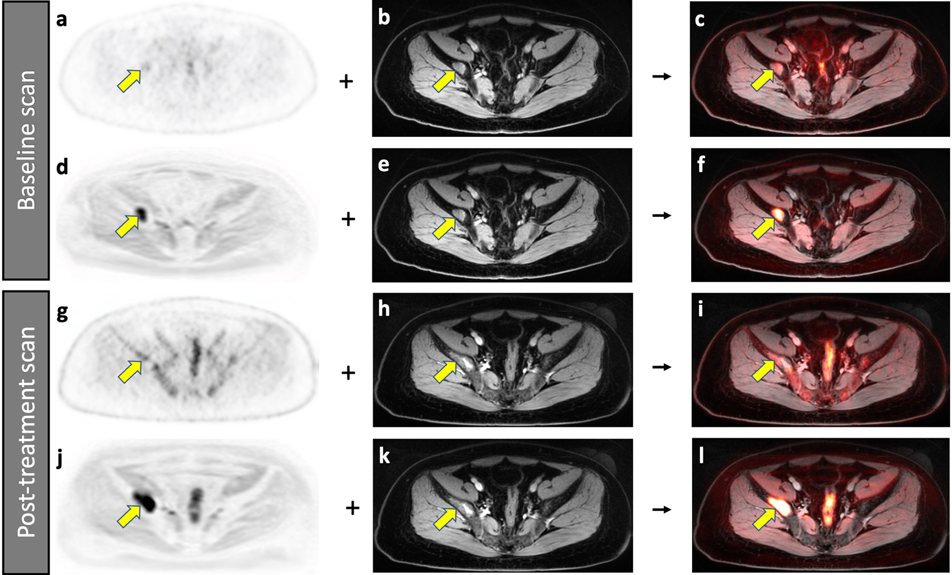
Improved detection of a bone marrow metastasis in an 18-year-old male patient with Ewing sarcoma on DW-MRI compared to 2-[18F]FDG-PET scan. Baseline scans (a-f), A bone marrow metastasis in the right iliac wing (arrow) demonstrates minor 2-[18F]FDG uptake on the PET scan (a). The lesion is also noted on the contrast-enhanced T1-weighted gradient echo scan (b) and the integrated 2-[18F]FDG-PET/MRI (c). The same bone marrow metastasis (arrow) demonstrates markedly restricted diffusion on the DW-MRI (d). After fusion with the contrast-enhanced MRI (e), it is better depicted on the integrated DW-MRI (f) than on the integrated 2-[18F]FDG-PET/MR scan. Follow up scans after 9 weeks of chemotherapy (g-l), On the post-treatment 2-[18F]FDG-PET scan (g), the normal bone marrow demonstrates increased hypermetabolic activity, which obscures the lesion (yellow arrow). The contrast-enhanced MRI demonstrates a larger area of inhomogeneous enhancement (h). It is difficult to determine a change in size or metabolic activity based on the integrated 2-[18F]FDG-PET/MRI (i). However, the DW-MRI scan (j) clearly demonstrates that the lesion increased in size. After fusion with the contrast-enhanced MRI (k), the integrated DW-MRI clearly demonstrates interval tumor growth (l). This case demonstrates that DW-MRI can sometimes better depict tumor progression than 2-[18F]FDG-PET.
