Figure 1.

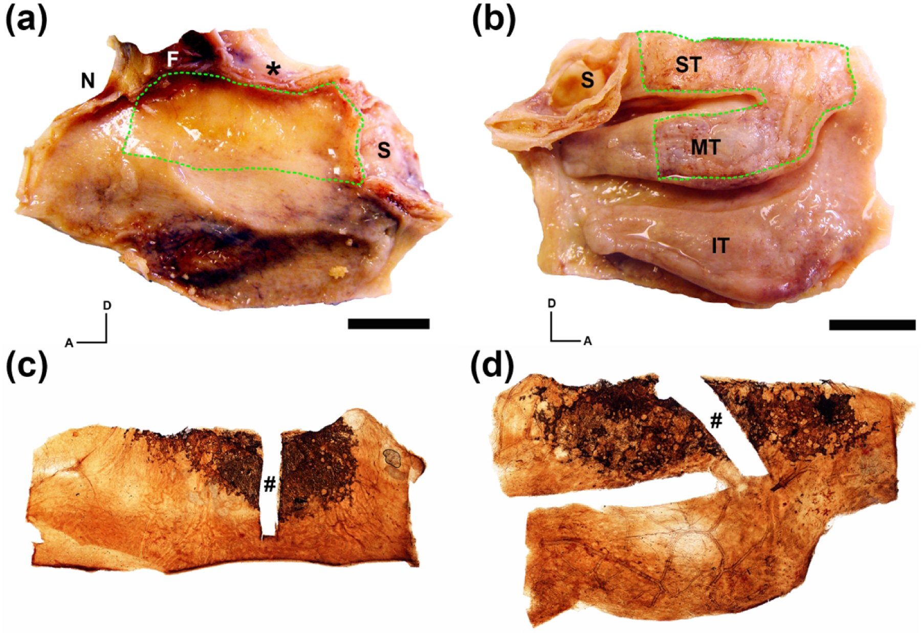
Generation of whole mucosal neuronal staining obtained from an 80 year-old male. The left nasal cavity autopsy specimen was split longitudinally along the cribriform plate (asterisk) exposing the septal surface (a) and lateral surface (b). (a, b). Whole mucosal sheets encompassing the region of olfactory mucosa were removed from the specimen (green dashed line). (c, d). Immunohistochemical staining of the excised septal (c) and lateral (d) mucosal sheets using anti-beta III tubulin (TUJ1) with DAB as a chromogen reveal brown neuronal staining of the olfactory receptor neurons at the dorsal portion of the specimens. A rectangular portion of the mucosal sheets used for sectioning and further histologic analysis was excised prior to whole mount staining (#). A = anterior; D = dorsal; F = frontal sinus; N = nasal bone; S = sphenoid sinus; IT = inferior turbinate; MT = middle turbinate; ST = superior turbinate; scale bars = 15 mm.
