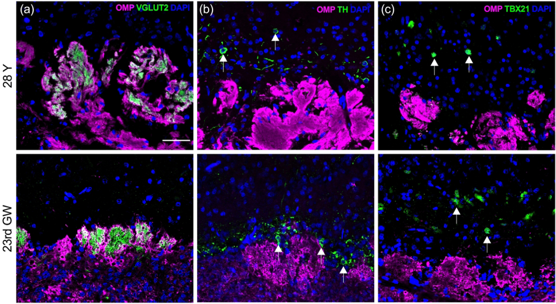
Figure 9.

OB glomeruli appear more disorganized with age. Near-adjacent coronal sections were obtained through the OB from a young-adult (#12-080313, 28 years old (28Y, male; top row) and embryo (#6167, 23rd gestation week (23rd GW), male; bottom row). (a). The OSN axon processes of the olfactory nerve layer at the periphery of the OB (oriented at the bottom of each panel) are labeled with antibodies to OMP (magenta). Terminal synapses with second order neurons are identified with antibodies against vesicular glutamate transporter type 2 (VGLUT2, green) within glomerular neuropil units. While fetal glomeruli appear as small buds just beginning to form into more robust structures, adult glomeruli are larger, more difficult to identify as discrete units, and often extend into deeper regions (see Fig 10(a) for lower magnification). (b). Adult periglomerular cells (arrows) labeled with antibodies against tyrosine hydroxylase (TH, green) are comparable in their position surrounding individual glomeruli in both adult and fetal tissue, but in embryonic tissue they appear closer to glomeruli. (c). Mitral cells (arrows) are labeled with anti-TBX21 antibodies (green). In adult OB they appear greatly reduced in number and somewhat sporadic while embryonic mitral cells demonstrate an established uniform layer located in a zone deep to the glomeruli as described in rodent OB. Scale bar = 50 μm.
