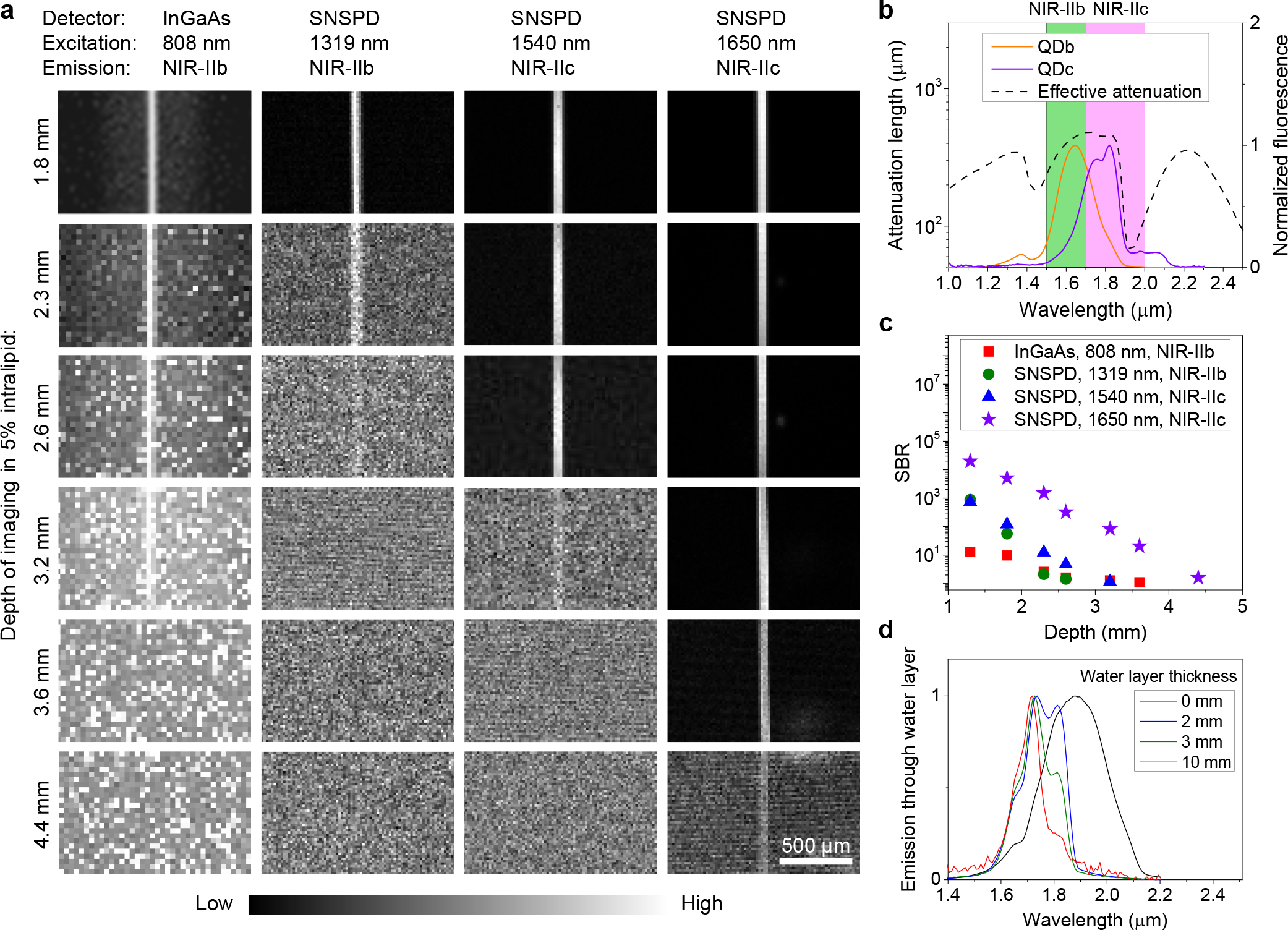
Figure 2 |. Fluorescence imaging in NIR-IIb and NIR-IIc windows.

(a) Fluorescence imaging of a 50-μm diameter capillary tube filled with NIR-IIb QDb or NIR-IIc QDc immersed at different depths in 5% intralipid by a wide-field system with a 2D InGaAs camera or a confocal microscope with SNSPDs. The 50-μm diameter capillary tubes immersed in 5% intralipid solution mimicked blood vessels in mouse brain tissues. An 808-nm laser was used for NIR-IIb wide-field imaging. A 1319 nm laser was applied for NIR-IIb confocal microscopy. A 1540-nm laser or a 1650-nm laser and a 10X objective (NA = 0.25) were used for NIR-IIc confocal microscopy. NIR-IIb and NIR-IIc fluorescence was collected in 1500–1700 nm or 1800–2000 nm ranges, respectively. These three lasers had the same power (28.5 mW) at intralipid surface. Similar results for n = 3 individual experiments. (b) Fluorescence spectra of P3-QDb and P3-QDc in PBS with emission peaks in NIR-IIb 12 and NIR-IIc regions respectively. A cuvette with 1-mm light path was used to measure the spectra. The effective attenuation length is from Fig. 1b. (c) Comparison of SBR of the results shown in (a). The SBR of wide-field results was from the ratio of the capillary brightness and the signal from sidelobe around the capillary. (d) Emission spectra of NIR-IIc QDc in a cuvette measured under water at depths of 2 mm, 3 mm and 10 mm respectively. The 0 mm data indicates no water layer and the emission spectrum was measured with QDc in tetrachloroethylene without any influence from water.
