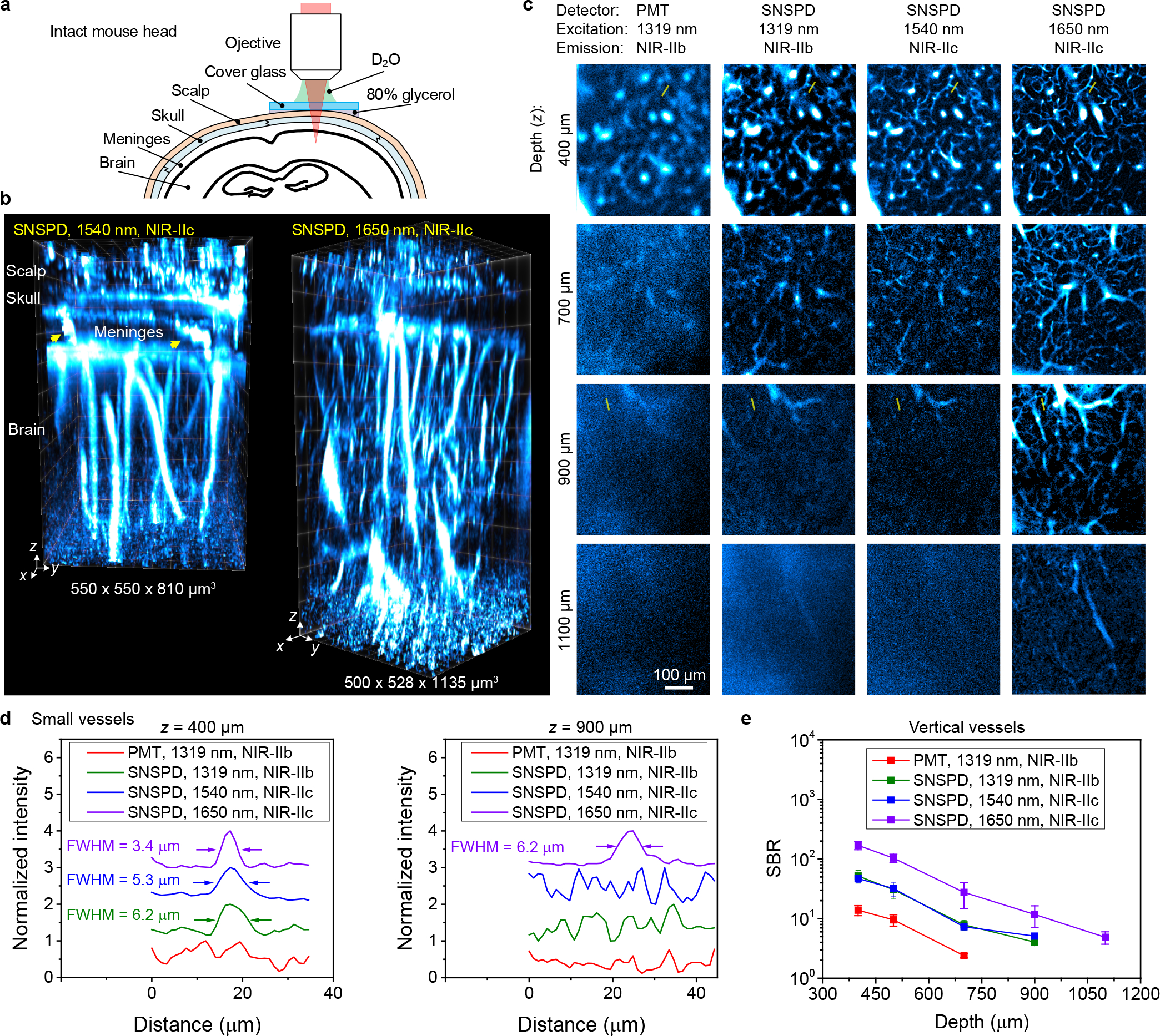
Figure 3 |. Non-invasive in vivo confocal microscopy of intact mouse head in NIR-IIc window.

(a) Schematic of intact mouse head imaging in NIR-IIc window. The gap between a ~170-μm thick cover glass and mouse head were filled with 80% glycerol. A 25X objective (NA = 1.05) and heavy water (D2O) as the immersion liquid were used. (b) 3D volumetric images of blood vessels in an intact mouse head visualized through the scalp, skull, meninges and brain cortex obtained with a 5-μm z-scan increment. The arrows point to the vessel like channels connecting the skull and brain cortex in the meninges. Confocal microscopy was performed 30 min after intravenous injection of P3-QDc. Left: fluorescence signal was collected in 1750–2000 nm window and excited by a 1540 nm laser. Right: a 1650 nm laser was used and fluorescence was collected in 1800–2000 nm window. (c) High-resolution confocal images of blood vessels at various depths through intact mouse head imaged with a PMT or SNSPD in NIR-IIb or NIR-IIc window. Imaging was performed 30 min after intravenous injection of P3-QDb and P3-QDc sequentially. NIR-IIb and NIR-IIc fluorescence was collected in 1500–1700 nm and 1800–2000 nm windows and excited by a 1319-nm, a 1540-nm laser and a 1650-nm laser, respectively. These three lasers had the same power (28.5 mW) at mouse head surface. (b,c) Similar results for n = 3 individuals (BALB/c, female, 3 weeks old). (d) Normalized photon counts profiles along the yellow lines in c. (e) Comparison of SBR for xy images recorded at different depths by confocal microscopy with a PMT or SNSPD in different windows. The signals of vertical vessels were used to calculate the SBR as these vessels existed from the surface of brain cortex to the deepest layer of imaging. Data are shown as mean ± s.d. derived from analyzing ≥ 4 vessels at each depth.
