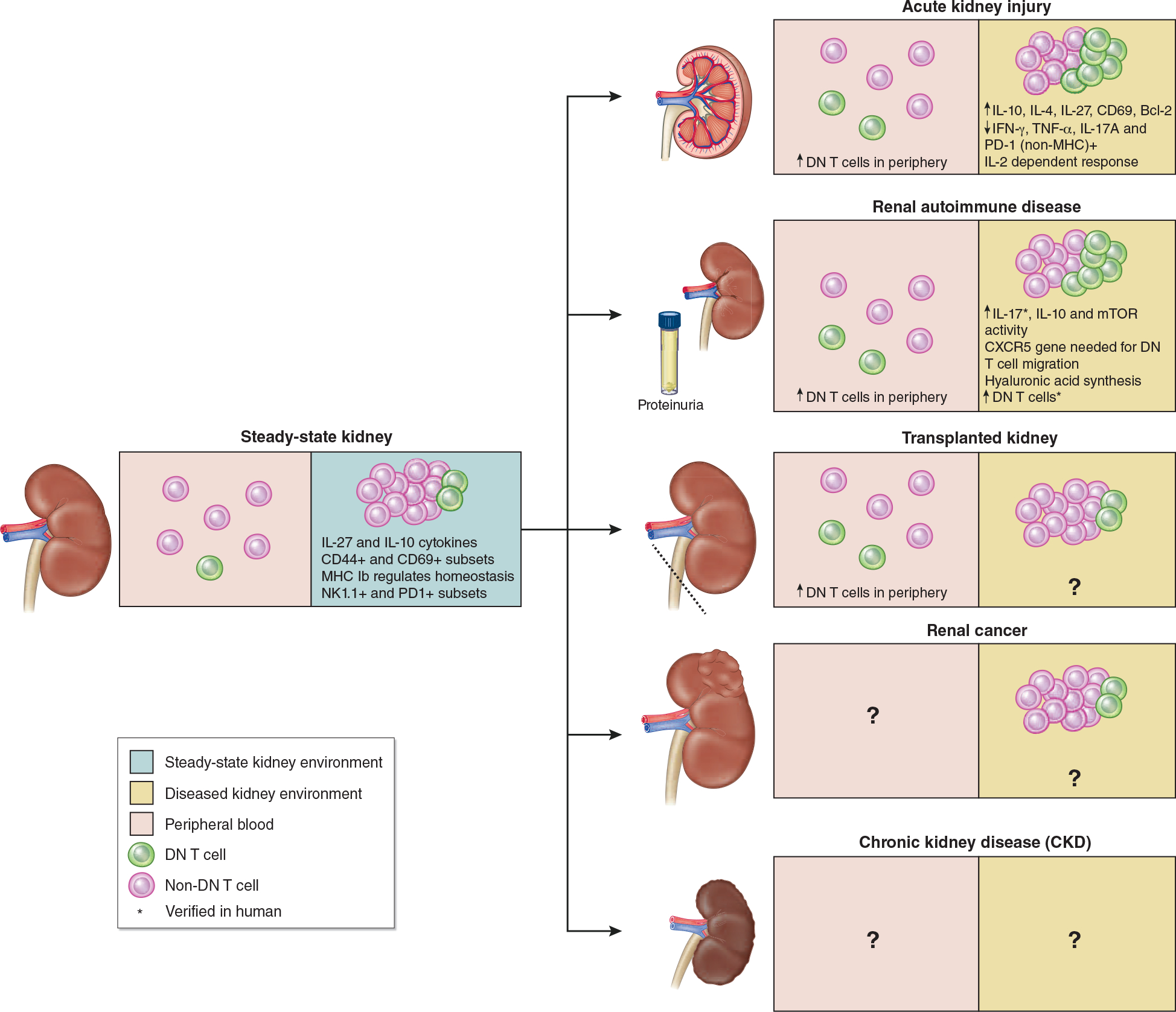
Figure 3. Role of DN T cells in different kidney disease models.

Note: DN T cells are in green and other αβT cells are in red. Proportion between DN T and other αβT cells are not proportional as DN T cells make up a very small percentage of αβT cells. Therefore, the increase in DN T cells is only for the purposes of showing general increases and decreases in DN T cell population percentage.
Steady-state DN T cells in kidney belong to 20–38% of αβT cells in normal murine kidney and show high frequency of IL-27 and IL-10 cytokines and CD44+ and CD69+ subsets.4 MHC Ib regulates homeostasis and two DN T cell subsets have been described – NK1.1+ and PD1+ DN T cells in murine and human kidney. Post-AKI kidney has shown increased DN T cell levels 24hr after injury both in kidney and periphery.4 Autoimmune kidney has shown increase in DN T cell population compared to steady-state kidney, however, DN T cells have been found to further contribute to autoimmune disease progression.31 Following transplanted kidney, peripheral DN T cells have been found to increase, yet the DN T cell population is unknown.19 However, DN T cell adoptive transfer has shown improved survival rates in other cardiac and skin allografts which leads to the hypothesis that this could improve survival rates of kidney transplants.32,33 DN T cell percentage in renal cancer is similar to adjacent tissue, however, the specific role of DN T cells in renal cancer remain unknown.34 We hypothesize treatment of renal cancer with DN T cell adoptive transfer could mitigate growth of cancerous cells.35 The same hypothesis can be made with regards to CKD since adoptive transfer of DN T cells to ischemic kidney has shown decreased kidney injury in mice and can therefore help mitigate AKI to CKD transition.5
